Roles of functional loops and the C-terminal segment of a single-stranded DNA binding protein elucidated by X-Ray structure analysis
Matsumoto, T., Morimoto, Y., Shibata, N., Kinebuchi, T., Shimamoto, N., Tsukihara, T., Yasuoka, N.(2000) J Biochem 127: 329-335
- PubMed: 10731701
- DOI: https://doi.org/10.1093/oxfordjournals.jbchem.a022611
- Primary Citation of Related Structures:
1EQQ, 1QVC - PubMed Abstract:
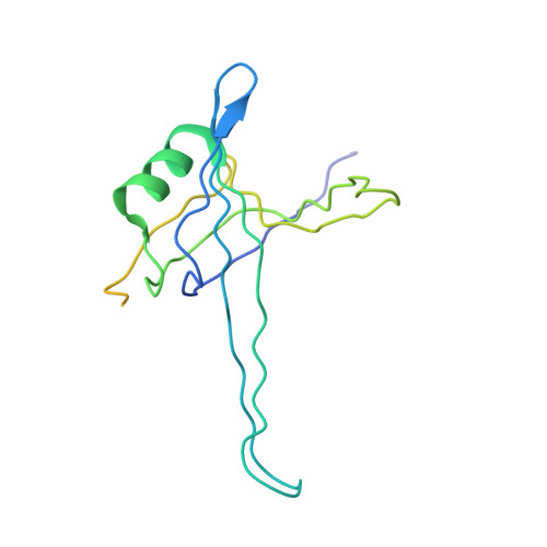
The single-stranded DNA (ssDNA) binding protein from Escherichia coli (EcoSSB) plays a central role in DNA replication, recombination and repair. The tertiary structure of EcoSSB was determined at 2.2 A resolution. This is rather higher resolution than previously reported. Crystals were grown from the homogeneous intact protein but the EcoSSB tetramer in the crystals contains truncated subunits lacking a part of the C-terminal. The structure determined includes biologically important flexible loops and C-terminal regions, and revealed the existence of concavities. These concavities include the residues important for ssDNA binding. An ssDNA can be fitted on the concavities and further stabilized through interactions with the loops forming flexible lids. It seems likely to play a central role in the binding of ssDNA.
Organizational Affiliation:
Department of Life Science, Himeji Institute of Technology, Kamigori, Ako-gun, Hyogo 678-1297, Japan.