


Structure of erythrocruorin in different ligand states refined at 1.4 A resolution.
Steigemann, W., Weber, E.(1979) J Mol Biol 127: 309-338
Experimental Data Snapshot
wwPDB Validation 3D Report Full Report

(1979) J Mol Biol 127: 309-338
Entity ID: 1 | |||||
|---|---|---|---|---|---|
| Molecule | Chains | Sequence Length | Organism | Details | Image |
| ERYTHROCRUORIN (CARBONMONOXY) | 136 | Chironomus thummi thummi | Mutation(s): 0 | 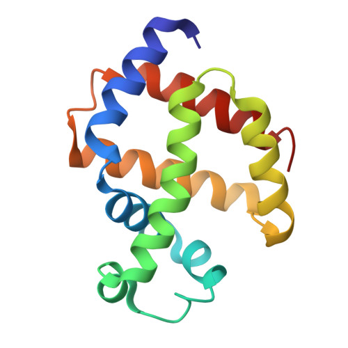 | |
UniProt | |||||
Find proteins for P02229 (Chironomus thummi thummi) Explore P02229 Go to UniProtKB: P02229 | |||||
Entity Groups | |||||
| Sequence Clusters | 30% Identity50% Identity70% Identity90% Identity95% Identity100% Identity | ||||
| UniProt Group | P02229 | ||||
Sequence AnnotationsExpand | |||||
| |||||
| Ligands 2 Unique | |||||
|---|---|---|---|---|---|
| ID | Chains | Name / Formula / InChI Key | 2D Diagram | 3D Interactions | |
| HEM Query on HEM | B [auth A] | PROTOPORPHYRIN IX CONTAINING FE C34 H32 Fe N4 O4 KABFMIBPWCXCRK-RGGAHWMASA-L |  | ||
| CMO Query on CMO | C [auth A] | CARBON MONOXIDE C O UGFAIRIUMAVXCW-UHFFFAOYSA-N |  | ||
| Length ( Å ) | Angle ( ˚ ) |
|---|---|
| a = 54.3 | α = 90 |
| b = 54.3 | β = 90 |
| c = 35.6 | γ = 120 |
RCSB PDB (citation) is hosted by
RCSB PDB is a member of the

