Cyclomaltodextrinase, Neopullulanase, and Maltogenic Amylase are Nearly Indistinguishable from Each Other
Lee, H.-S., Kim, M.-S., Cho, H.-S., Kim, J.-I., Kim, T.-J., Choi, J.-H., Park, C., Lee, H.-S., Oh, B.-H., Park, K.-H.(2002) J Biol Chem 277: 21891
- PubMed: 11923309
- DOI: https://doi.org/10.1074/jbc.M201623200
- Primary Citation of Related Structures:
1EA9, 1GVI - PubMed Abstract:
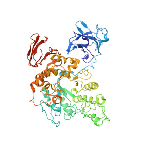
Over 20 enzymes denoted as cyclomaltodextrinase, maltogenic amylase, or neopullulanase that share 40-86% sequence identity with each other are found in public data bases. These enzymes are distinguished from typical alpha-amylases by containing a novel N-terminal domain and exhibiting preferential substrate specificities for cyclomaltodextrins (CDs) over starch. In this research field, a great deal of confusion exists regarding the features distinguishing the three groups of enzymes from one another. Although a different enzyme code has been assigned to each of the three different enzyme names, even a single differentiating enzymatic property has not been documented in the literature. On the other hand, an outstanding question related to this issue concerns the structural basis for the preference of these enzymes for CDs. To clarify the confusion and to address this question, we have determined the structures of two enzymes, one from alkalophilic Bacillus sp. I-5 and named cyclomaltodextrinase and the other from a Thermus species and named maltogenic amylase. The structure of the Bacillus enzyme reveals a dodecameric assembly composed of six copies of the dimer, which is the structural and functional unit of the Thermus enzyme and an enzyme named neopullulanase. The structure of the Thermus enzyme in complex with beta-CD led to the conclusion that Trp47, a well conserved N-terminal domain residue, contributes greatly to the preference for beta-CD. The common dimer formation through the novel N-terminal domain, which contributes to the preference for CDs by lining the active-site cavity, convincingly indicates that the three groups of enzymes are not different enough to preserve the different names and enzyme codes.
Organizational Affiliation:
National Creative Research Initiative Center for Biomolecular Recognition, Department of Life Science, Pohang University of Science and Technology, Pohang, Kyungbuk 790-784, Korea.