Reversible Dissociation of Thiolate Ligands from Molybdenum in an Enzyme of the Dimethyl Sulfoxide Reductase Family
Bray, R.C., Adams, B., Smith, A.T., Bennett, B., Bailey, S.(2000) Biochemistry 39: 11258
- PubMed: 10985771
- DOI: https://doi.org/10.1021/bi0000521
- Primary Citation of Related Structures:
1E5V, 1E60, 1E61 - PubMed Abstract:
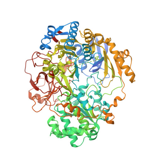
Much is unknown concerning the role of thiolate ligands of molybdenum in molybdopterin enzymes. It has been suggested that thiolate dissociation from molybdenum is part of the catalytic mechanism of bis-molybdopterin enzymes of the dimethyl sulfoxide reductase (DMSOR) family. For DMSOR from Rhodobacter capsulatus, thiolate dissociation has therefore been investigated crystallographically, by UV/visible spectroscopy, and by enzyme assays. When crystallized from sodium citrate, all four thiolates of DMSOR are within bonding distance of Mo, but after extended exposure to Na(+)-Hepes, a pair of thiolates dissociates, a mixture of structures being indicated after shorter exposures to this buffer. DMSOR is stable in sodium citrate and other buffers but unstable aerobically although not anaerobically in Na(+)-Hepes. Aerobically in Na(+)-Hepes, a first-order reaction (k = 0.032 hr(-)(1) at 37 degrees C) leads to loss of activity in the backward but not the forward (dimethyl sulfoxide reduction) assay and loss of absorption at lambda > approximately 450 nm. This reaction can be reversed by a cycle of reduction and reoxidation ("redox-cycling"). Slower irreversible loss of activity in the forward assay and cofactor dissociation follow. Spectral analogy with a mono-molybdopterin enzyme supports the conclusion that in the Hepes-modified DMSOR form, only two cofactor dithiolene sulfur atoms are coordinated to molybdenum. Loss of activity provides the first clear evidence that sulfur ligand dissociation is an artifact, not part of the catalytic cycle. Clearly, structural data on DMSOR samples extensively exposed to Hepes is not directly relevant to the native enzyme. The nature of the oxygen ligands detected crystallographically is discussed, as is the specificity of Hepes and the mechanism whereby its effects are achieved. DMSOR forms complexes with Na(+)-Hepes and other buffer ions. For DMSOR crystallized from Hepes, electron density in the substrate binding channel suggests that buffers bind in this site. Like the as-prepared enzyme, the modified form (DMSOR(mod)D), known to arise on extended aerobic exposure to dimethyl sulfide, is susceptible to a further degradative reaction, although this is not buffer-dependent. It involves loss of absorption at lambda > approximately 450 nm and, presumably, dissociation of thiolate ligands. Evidence is presented that, as a result of O(2) damage, DMSOR samples not submitted to redox-cycling may be contaminated with DMSOR(mod)D and with material absorbing in the region of 400 nm, analogous to the Hepes-modified enzyme. Since the latter lacks absorption at lambda > approximately 450 nm, its presence may escape detection.
Organizational Affiliation:
School of Chemistry, Physics and Environmental Science, University of Sussex, Brighton, BN1 9QJ, UK.