1E39

Flavocytochrome C3 from Shewanella frigidimarina histidine 365 mutated to alanine


Experimental Data Snapshot

  • Method: X-RAY DIFFRACTION
  • Resolution: 1.80 Å
  • R-Value Free: 0.247 
  • R-Value Observed: 0.181 

wwPDB Validation   3D Report Full Report


This is version 2.1 of the entry. See complete history


Literature

Identification of the Active Site Acid/Base Catalyst in a Bacterial Fumarate Reductase: A Kinetic and Crystallographic Study

Doherty, M.K.Pealing, S.L.Miles, C.S.Moysey, R.Taylor, P.Walkinshaw, M.D.Reid, G.A.Chapman, S.K.

(2000) Biochemistry 39: 10695

  • DOI: https://doi.org/10.1021/bi000871l
  • Primary Citation of Related Structures:  
    1E39

  • PubMed Abstract: 

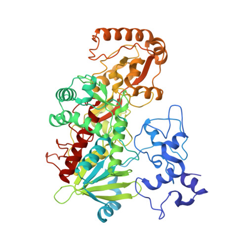
    The active sites of respiratory fumarate reductases are highly conserved, indicating a common mechanism of action involving hydride and proton transfer. Evidence from the X-ray structures of substrate-bound fumarate reductases, including that for the enzyme from Shewanella frigidimarina [Taylor, P., Pealing, S. L., Reid, G. A., Chapman, S. K., and Walkinshaw, M. D. (1999) Nat. Struct. Biol. 6, 1108-1112], indicates that the substrate is well positioned to accept a hydride from N5 of the FAD. However, the identity of the proton donor has been the subject of recent debate and has been variously proposed to be (using numbering for the S. frigidimarina enzyme) His365, His504, and Arg402. We have used site-directed mutagenesis to examine the roles of these residues in the S. frigidimarina enzyme. The H365A and H504A mutant enzymes exhibited lower k(cat) values than the wild-type enzyme but only by factors of 3-15, depending on pH. This, coupled with the increase in K(m) observed for these enzymes, indicates that His365 and His504 are involved in Michaelis complex formation and are not essential catalytic residues. In fact, examination of the crystal structure of S. frigidimarina fumarate reductase has led to the proposal that Arg402 is the only plausible active site acid. Consistent with this proposal, we report that the R402A mutant enzyme has no detectable fumarate reductase activity. The crystal structure of the H365A mutant enzyme shows that, in addition to the replacement at position 365, there have been some adjustments in the positions of active site residues. In particular, the observed change in the orientation of the Arg402 side chain could account for the decrease in k(cat) seen with the H365A enzyme. These results demonstrate that an active site arginine and not a histidine residue is the proton donor for fumarate reduction.


  • Organizational Affiliation

    Department of Chemistry, University of Edinburgh, West Mains Road, Edinburgh EH9 3JJ, U.K.


Macromolecules
Find similar proteins by:  (by identity cutoff)  |  3D Structure
Entity ID: 1
MoleculeChains Sequence LengthOrganismDetailsImage
FUMARATE REDUCTASE FLAVOPROTEIN SUBUNIT571Shewanella frigidimarinaMutation(s): 1 
EC: 1.3.99.1
UniProt
Find proteins for P0C278 (Shewanella frigidimarina)
Explore P0C278 
Go to UniProtKB:  P0C278
Entity Groups  
Sequence Clusters30% Identity50% Identity70% Identity90% Identity95% Identity100% Identity
UniProt GroupP0C278
Sequence Annotations
Expand
  • Reference Sequence
Small Molecules
Ligands 5 Unique
IDChains Name / Formula / InChI Key2D Diagram3D Interactions
FAD
Query on FAD

Download Ideal Coordinates CCD File 
F [auth A]FLAVIN-ADENINE DINUCLEOTIDE
C27 H33 N9 O15 P2
VWWQXMAJTJZDQX-UYBVJOGSSA-N
HEC
Query on HEC

Download Ideal Coordinates CCD File 
B [auth A],
C [auth A],
D [auth A],
E [auth A]
HEME C
C34 H34 Fe N4 O4
HXQIYSLZKNYNMH-LJNAALQVSA-N
FUM
Query on FUM

Download Ideal Coordinates CCD File 
G [auth A]FUMARIC ACID
C4 H4 O4
VZCYOOQTPOCHFL-OWOJBTEDSA-N
GOL
Query on GOL

Download Ideal Coordinates CCD File 
I [auth A]GLYCEROL
C3 H8 O3
PEDCQBHIVMGVHV-UHFFFAOYSA-N
NA
Query on NA

Download Ideal Coordinates CCD File 
H [auth A]SODIUM ION
Na
FKNQFGJONOIPTF-UHFFFAOYSA-N
Experimental Data & Validation

Experimental Data

  • Method: X-RAY DIFFRACTION
  • Resolution: 1.80 Å
  • R-Value Free: 0.247 
  • R-Value Observed: 0.181 
  • Space Group: P 1 21 1
Unit Cell:
Length ( Å )Angle ( ˚ )
a = 45.571α = 90
b = 92.172β = 91.08
c = 78.489γ = 90
Software Package:
Software NamePurpose
SHELXL-97refinement
DENZOdata reduction
SCALEPACKdata scaling
SHELXL-97phasing

Structure Validation

View Full Validation Report


Entry History 

Deposition Data

Revision History  (Full details and data files)

  • Version 1.0: 2000-09-18
    Type: Initial release
  • Version 1.1: 2011-08-24
    Changes: Database references, Derived calculations, Non-polymer description, Other, Structure summary, Version format compliance
  • Version 1.2: 2019-05-08
    Changes: Data collection, Derived calculations, Experimental preparation, Other
  • Version 1.3: 2019-05-22
    Changes: Data collection, Refinement description
  • Version 2.0: 2023-11-15
    Changes: Atomic model, Data collection, Database references, Derived calculations
  • Version 2.1: 2023-12-06
    Changes: Refinement description