1DGJ

CRYSTAL STRUCTURE OF THE ALDEHYDE OXIDOREDUCTASE FROM DESULFOVIBRIO DESULFURICANS ATCC 27774


Experimental Data Snapshot

  • Method: X-RAY DIFFRACTION
  • Resolution: 2.80 Å
  • R-Value Free: 0.224 
  • R-Value Work: 0.164 
  • R-Value Observed: 0.167 

wwPDB Validation   3D Report Full Report


This is version 1.3 of the entry. See complete history


Literature

Gene sequence and crystal structure of the aldehyde oxidoreductase from Desulfovibrio desulfuricans ATCC 27774.

Rebelo, J.Macieira, S.Dias, J.M.Huber, R.Ascenso, C.S.Rusnak, F.Moura, J.J.Moura, I.Romao, M.J.

(2000) J Mol Biol 297: 135-146

  • DOI: https://doi.org/10.1006/jmbi.2000.3552
  • Primary Citation of Related Structures:  
    1DGJ

  • PubMed Abstract: 

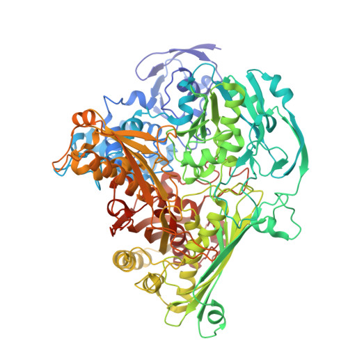
    The aldehyde oxidoreductase (MOD) isolated from the sulfate reducer Desulfovibrio desulfuricans (ATCC 27774) is a member of the xanthine oxidase family of molybdenum-containing enzymes. It has substrate specificity similar to that of the homologous enzyme from Desulfovibrio gigas (MOP) and the primary sequences from both enzymes show 68 % identity. The enzyme was crystallized in space group P6(1)22, with unit cell dimensions of a=b=156.4 A and c=177.1 A, and diffraction data were obtained to beyond 2.8 A. The crystal structure was solved by Patterson search techniques using the coordinates of the D. gigas enzyme. The overall fold of the D. desulfuricans enzyme is very similar to MOP and the few differences are mapped to exposed regions of the molecule. This is reflected in the electrostatic potential surfaces of both homologous enzymes, one exception being the surface potential in a region identifiable as the putative docking site of the physiological electron acceptor. Other essential features of the MOP structure, such as residues of the active-site cavity, are basically conserved in MOD. Two mutations are located in the pocket bearing a chain of catalytically relevant water molecules. As deduced from this work, both these enzymes are very closely related in terms of their sequences as well as 3D structures. The comparison allowed confirmation and establishment of features that are essential for their function; namely, conserved residues in the active-site, catalytically relevant water molecules and recognition of the physiological electron acceptor docking site.


  • Organizational Affiliation

    Departamento de Química Centro de Química Fina e Biotecnologia, Faculdade de Ciências e Tecnologia, Universidade Nova de Lisboa, Caparica, Portugal.


Macromolecules
Find similar proteins by:  (by identity cutoff)  |  3D Structure
Entity ID: 1
MoleculeChains Sequence LengthOrganismDetailsImage
ALDEHYDE OXIDOREDUCTASE907Desulfovibrio desulfuricansMutation(s): 0 
EC: 1.2
UniProt
Find proteins for Q9REC4 (Desulfovibrio desulfuricans)
Explore Q9REC4 
Go to UniProtKB:  Q9REC4
Entity Groups  
Sequence Clusters30% Identity50% Identity70% Identity90% Identity95% Identity100% Identity
UniProt GroupQ9REC4
Sequence Annotations
Expand
  • Reference Sequence
Small Molecules
Ligands 3 Unique
IDChains Name / Formula / InChI Key2D Diagram3D Interactions
MCN
Query on MCN

Download Ideal Coordinates CCD File 
E [auth A]PTERIN CYTOSINE DINUCLEOTIDE
C19 H22 N8 O13 P2 S2
RBWYFPNWTRZKKZ-LOIMWUFNSA-N
FES
Query on FES

Download Ideal Coordinates CCD File 
B [auth A],
C [auth A]
FE2/S2 (INORGANIC) CLUSTER
Fe2 S2
NIXDOXVAJZFRNF-UHFFFAOYSA-N
2MO
Query on 2MO

Download Ideal Coordinates CCD File 
D [auth A]MOLYBDENUM (IV)OXIDE
Mo O2
QXYJCZRRLLQGCR-UHFFFAOYSA-N
Experimental Data & Validation

Experimental Data

  • Method: X-RAY DIFFRACTION
  • Resolution: 2.80 Å
  • R-Value Free: 0.224 
  • R-Value Work: 0.164 
  • R-Value Observed: 0.167 
  • Space Group: P 61 2 2
Unit Cell:
Length ( Å )Angle ( ˚ )
a = 156.639α = 90
b = 156.639β = 90
c = 177.149γ = 120
Software Package:
Software NamePurpose
AMoREphasing
X-PLORrefinement
DENZOdata reduction
SCALEPACKdata scaling

Structure Validation

View Full Validation Report


Entry History 

Deposition Data

Revision History  (Full details and data files)

  • Version 1.0: 2000-03-22
    Type: Initial release
  • Version 1.1: 2008-04-27
    Changes: Version format compliance
  • Version 1.2: 2011-07-13
    Changes: Version format compliance
  • Version 1.3: 2024-02-07
    Changes: Data collection, Database references, Derived calculations