1D3H

HUMAN DIHYDROOROTATE DEHYDROGENASE COMPLEXED WITH ANTIPROLIFERATIVE AGENT A771726


Experimental Data Snapshot

  • Method: X-RAY DIFFRACTION
  • Resolution: 1.80 Å
  • R-Value Free: 0.185 
  • R-Value Work: 0.162 
  • R-Value Observed: 0.162 

wwPDB Validation   3D Report Full Report


Ligand Structure Quality Assessment 


This is version 1.4 of the entry. See complete history


Literature

Structures of human dihydroorotate dehydrogenase in complex with antiproliferative agents.

Liu, S.Neidhardt, E.A.Grossman, T.H.Ocain, T.Clardy, J.

(2000) Structure 8: 25-33

  • DOI: https://doi.org/10.1016/s0969-2126(00)00077-0
  • Primary Citation of Related Structures:  
    1D3G, 1D3H

  • PubMed Abstract: 

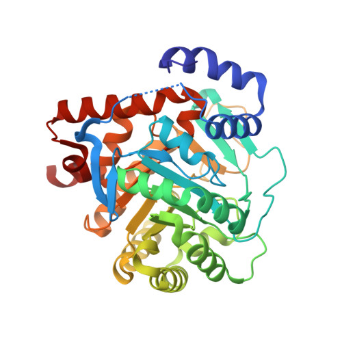
    Dihydroorotate dehydrogenase (DHODH) catalyzes the fourth committed step in the de novo biosynthesis of pyrimidines. As rapidly proliferating human T cells have an exceptional requirement for de novo pyrimidine biosynthesis, small molecule DHODH inhibitors constitute an attractive therapeutic approach to autoimmune diseases, immunosuppression, and cancer. Neither the structure of human DHODH nor any member of its family was known. The high-resolution crystal structures of human DHODH in complex with two different inhibitors have been solved. The initial set of phases was obtained using multiwavelength anomalous diffraction phasing with selenomethionine-containing DHODH. The structures have been refined to crystallographic R factors of 16.8% and 16.2% at resolutions of 1. 6 A and 1.8 A for inhibitors related to brequinar and leflunomide, respectively. Human DHODH has two domains: an alpha/beta-barrel domain containing the active site and an alpha-helical domain that forms the opening of a tunnel leading to the active site. Both inhibitors share a common binding site in this tunnel, and differences in the binding region govern drug sensitivity or resistance. The active site of human DHODH is generally similar to that of the previously reported bacterial active site. The greatest differences are that the catalytic base removing the proton from dihydroorotate is a serine rather than a cysteine, and that packing of the flavin mononucleotide in its binding site is tighter.


  • Organizational Affiliation

    Department of Chemistry and Chemical Biology, Cornell University, Ithaca, NY 14853-1301, USA.


Macromolecules
Find similar proteins by:  (by identity cutoff)  |  3D Structure
Entity ID: 1
MoleculeChains Sequence LengthOrganismDetailsImage
DIHYDROOROTATE DEHYDROGENASE367Homo sapiensMutation(s): 0 
EC: 1.3.3.1
Membrane Entity: Yes 
UniProt & NIH Common Fund Data Resources
Find proteins for Q02127 (Homo sapiens)
Explore Q02127 
Go to UniProtKB:  Q02127
PHAROS:  Q02127
GTEx:  ENSG00000102967 
Entity Groups  
Sequence Clusters30% Identity50% Identity70% Identity90% Identity95% Identity100% Identity
UniProt GroupQ02127
Sequence Annotations
Expand
  • Reference Sequence
Small Molecules
Ligands 5 Unique
IDChains Name / Formula / InChI Key2D Diagram3D Interactions
FMN
Query on FMN

Download Ideal Coordinates CCD File 
E [auth A]FLAVIN MONONUCLEOTIDE
C17 H21 N4 O9 P
FVTCRASFADXXNN-SCRDCRAPSA-N
A26
Query on A26

Download Ideal Coordinates CCD File 
D [auth A](2Z)-2-cyano-3-hydroxy-N-[4-(trifluoromethyl)phenyl]but-2-enamide
C12 H9 F3 N2 O2
UTNUDOFZCWSZMS-YFHOEESVSA-N
ORO
Query on ORO

Download Ideal Coordinates CCD File 
F [auth A]OROTIC ACID
C5 H4 N2 O4
PXQPEWDEAKTCGB-UHFFFAOYSA-N
SO4
Query on SO4

Download Ideal Coordinates CCD File 
B [auth A]SULFATE ION
O4 S
QAOWNCQODCNURD-UHFFFAOYSA-L
ACT
Query on ACT

Download Ideal Coordinates CCD File 
C [auth A]ACETATE ION
C2 H3 O2
QTBSBXVTEAMEQO-UHFFFAOYSA-M
Binding Affinity Annotations 
IDSourceBinding Affinity
A26 BindingDB:  1D3H IC50: min: 17, max: 5012 (nM) from 15 assay(s)
EC50: min: 8900, max: 8913 (nM) from 2 assay(s)
Experimental Data & Validation

Experimental Data

  • Method: X-RAY DIFFRACTION
  • Resolution: 1.80 Å
  • R-Value Free: 0.185 
  • R-Value Work: 0.162 
  • R-Value Observed: 0.162 
  • Space Group: P 32 2 1
Unit Cell:
Length ( Å )Angle ( ˚ )
a = 90.61α = 90
b = 90.61β = 90
c = 122.41γ = 120
Software Package:
Software NamePurpose
MOSFLMdata reduction
SCALAdata scaling
SHELXSphasing
CNSrefinement
CCP4data scaling

Structure Validation

View Full Validation Report


Ligand Structure Quality Assessment 


Entry History 

Deposition Data

Revision History  (Full details and data files)

  • Version 1.0: 2000-08-13
    Type: Initial release
  • Version 1.1: 2008-04-27
    Changes: Version format compliance
  • Version 1.2: 2011-07-13
    Changes: Version format compliance
  • Version 1.3: 2012-07-18
    Changes: Non-polymer description
  • Version 1.4: 2024-02-07
    Changes: Data collection, Database references, Derived calculations