Influence of aglycone modifications on the binding of anthracycline drugs to DNA: the molecular structure of idarubicin and 4-O-demethyl-11-deoxydoxorubicin complexed to d(CGATCG).
Gao, Y.G., Wang, A.H.(1991) Anticancer Drug Des 6: 137-149
- PubMed: 1872945
- Primary Citation of Related Structures:
1D37, 1D38 - PubMed Abstract:
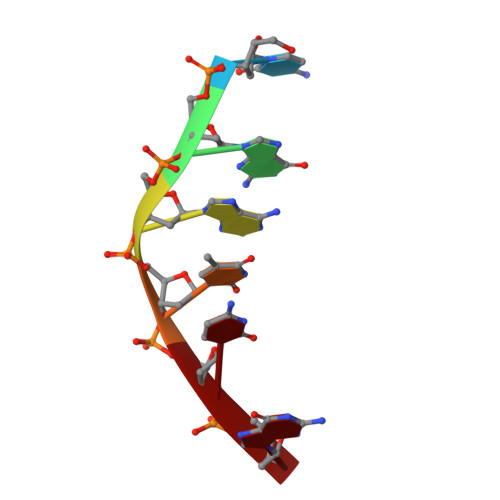
X-ray diffraction analyses of the complexes between two anthracycline antitumor compounds, idarubicin (IDR) and 4-O-demethyl-11-deoxydoxorubicin (ddDOX), with the DNA hexamer d(CGATCG) provided the detailed three-dimensional molecular structures at 1.7 A and 1.8 A resolution, respectively. Their structures have been refined with the constrained refinement procedure to final R-factors of 0.188 (1724 reflections for IDR) and 0.179 (1247 reflections for ddDOX). The overall structures of both complexes are similar to those of the previously studied DAU- and DOX-DNA complexes. In both complexes, two IDR (and ddDOX) molecules bind to the DNA hexamer double helix with the elongated aglycone chromophore intercalated between the CpG steps at both ends of the helix. The aglycone chromophore spans the GC Watson-Crick base pairs with its amino sugar lying in the minor groove where little structural difference is seen, compared with the daunorubicin-d(CGATCG) and doxorubicin-d(CGATCG) complexes. In contrast, the missing C4 methoxy of IDR and the missing methyl group at the O4 position of ddDOX result in a different binding surface in the major groove. The O4 hydroxyl group is capable of receiving and/or donating a hydrogen bond to proteins that bind to the drug-DNA complex. The missing O11 hydroxyl group in ring B creates an empty space in the intercalation cavity between the two GC base pairs, which appears to affect the stacking interactions between the aglycone and the DNA base pairs. Those structural changes in the major groove of the drug-DNA complexes due to the modifications of the aglycone chromophore may be responsible in part for the difference in their biological activities.
Organizational Affiliation:
Department of Physiology and Biophysics, University of Illinois, Urbana 61801.