1CZI

CHYMOSIN COMPLEX WITH THE INHIBITOR CP-113972


Experimental Data Snapshot

  • Method: X-RAY DIFFRACTION
  • Resolution: 2.30 Å
  • R-Value Work: 0.195 

wwPDB Validation   3D Report Full Report


This is version 3.0 of the entry. See complete history


Literature

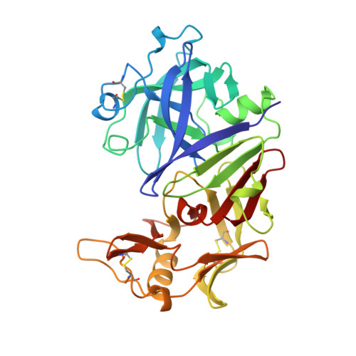
A 2.3 A resolution structure of chymosin complexed with a reduced bond inhibitor shows that the active site beta-hairpin flap is rearranged when compared with the native crystal structure.

Groves, M.R.Dhanaraj, V.Badasso, M.Nugent, P.Pitts, J.E.Hoover, D.J.Blundell, T.L.

(1998) Protein Eng 11: 833-840

  • DOI: https://doi.org/10.1093/protein/11.10.833
  • Primary Citation of Related Structures:  
    1CZI

  • PubMed Abstract: 

    In the crystal structure of uncomplexed native chymosin, the beta-hairpin at the active site, known as 'the flap', adopts a different conformation from that of other aspartic proteinases. This conformation would prevent the mode of binding of substrates/inhibitors generally found in other aspartic proteinase complexes. We now report the X-ray analysis of chymosin complexed with a reduced bond inhibitor CP-113972 ¿(2R,3S)-isopropyl 3-[(L-prolyl-p-iodo-L-phenylalanyl-S-methyl-cysteinyl)amino-4]-cyclohexy l-2-hydroxybutanoate¿ at 2.3 A resolution in a novel crystal form of spacegroup R32. The structure has been refined by restrained least-squares methods to a final R-factor of 0.19 for a total of 11 988 independent reflections in the resolution range 10 to 2.3 A. The extended beta-strand conformation of the inhibitor allows hydrogen bonds within the active site, while its sidechains make both electrostatic and hydrophobic interactions with residues lining the specificity pockets S4-->S1. The flap closes over the active site cleft in a way that closely resembles that of other previously determined aspartic proteinase inhibitor complexes. We conclude that the usual position and conformation of the flap found in other aspartic proteinases is available to native chymosin. The conformation observed in the native crystal form may result from intermolecular interactions between symmetry-related molecules in the crystal lattice.


  • Organizational Affiliation

    Department of Crystallography, Birkbeck College, London, UK.


Macromolecules
Find similar proteins by:  (by identity cutoff)  |  3D Structure
Entity ID: 1
MoleculeChains Sequence LengthOrganismDetailsImage
CHYMOSINA [auth E]323Bos taurusMutation(s): 0 
EC: 3.4.23.4
UniProt
Find proteins for P00794 (Bos taurus)
Explore P00794 
Go to UniProtKB:  P00794
Entity Groups  
Sequence Clusters30% Identity50% Identity70% Identity90% Identity95% Identity100% Identity
UniProt GroupP00794
Sequence Annotations
Expand
  • Reference Sequence

Find similar proteins by:  Sequence   |   3D Structure  

Entity ID: 2
MoleculeChains Sequence LengthOrganismDetailsImage
CP-113972 (NORSTATINE-S-METHYL CYSTEINE-IODO-PHENYLALANINE-PROLINE)B [auth P]4N/AMutation(s): 0 
Sequence Annotations
Expand
  • Reference Sequence
Small Molecules
Modified Residues  2 Unique
IDChains TypeFormula2D DiagramParent
PHI
Query on PHI
B [auth P]L-PEPTIDE LINKINGC9 H10 I N O2PHE
SMC
Query on SMC
B [auth P]L-PEPTIDE LINKINGC4 H9 N O2 SCYS
Biologically Interesting Molecules (External Reference) 1 Unique
Experimental Data & Validation

Experimental Data

  • Method: X-RAY DIFFRACTION
  • Resolution: 2.30 Å
  • R-Value Work: 0.195 
  • Space Group: H 3 2
Unit Cell:
Length ( Å )Angle ( ˚ )
a = 132.78α = 90
b = 132.78β = 90
c = 81.95γ = 120
Software Package:
Software NamePurpose
MOSFLMdata reduction
AGROVATA/ROTAVATAdata reduction
AMoREphasing
RESTRAINrefinement
CCP4data scaling
ROTAVATAdata scaling

Structure Validation

View Full Validation Report


Entry History 

Deposition Data

Revision History  (Full details and data files)

  • Version 1.0: 1997-04-01
    Type: Initial release
  • Version 1.1: 2008-03-24
    Changes: Version format compliance
  • Version 1.2: 2011-07-13
    Changes: Atomic model, Database references, Derived calculations, Non-polymer description, Structure summary, Version format compliance
  • Version 1.3: 2012-12-12
    Changes: Other
  • Version 2.0: 2023-08-09
    Changes: Atomic model, Database references, Derived calculations, Refinement description
  • Version 3.0: 2023-11-15
    Changes: Advisory, Atomic model, Data collection, Derived calculations