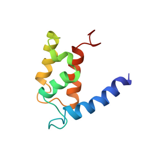
The solution structure of the bovine S100B protein dimer in the calcium-free state.
Kilby, P.M., Van Eldik, L.J., Roberts, G.C.(1996) Structure 4: 1041-1052
- PubMed: 8805590
- DOI: https://doi.org/10.1016/s0969-2126(96)00111-6
- Primary Citation of Related Structures:
1CFP - PubMed Abstract:
S100B (S100beta) is a member of the S100 family of small calcium-binding proteins: members of this family contain two helix-loop-helix calcium-binding motifs and interact with a wide range of proteins involved mainly in the cytoskeleton and cell proliferation. S100B is a neurite-extension factor and levels of S100B are elevated in the brains of patients with Alzheimer's disease or Down's syndrome: the pattern of S100B overexpression in Alzheimer's disease correlates with the pattern of neuritic-plaque formation. Identification of a growing class of S100 proteins and the likely neurochemical importance of S100B make the determination of the structure of S100B of interest. We have used NMR to determine the structure of the reduced S100B homodimer in the absence of calcium. Each monomer consists of a four-helix bundle, arranged in the dimer in an antiparallel fashion. The fourth helix of each monomer runs close to the equivalent helix of the other monomer for almost its full length, extending the hydrophobic core through the interface. The N-terminal, but not the C-terminal, calcium-binding loop is similar to the equivalent loop in the monomeric S100 protein calbindin and is in a conformation ready to bind calcium. The novel dimer structure reported previously for calcyclin (S100A6) is the common fold for the dimeric S100B proteins. Calcium binding to the C-terminal calcium-binding loop would be expected to require a conformational change, which might provide a signal for activation. The structure suggests regions of the molecule likely to be involved in interactions with effector molecules.
Organizational Affiliation:
Department of Biochemistry and Biological NMR Centre, Adrian Building, University of Leicester, Leicester, LE1 7RH, UK. pmk@le.ac.uk