1BWA

HIV-1 PROTEASE (V82F/I84V) DOUBLE MUTANT COMPLEXED WITH XV638 OF DUPONT PHARMACEUTICALS


Experimental Data Snapshot

  • Method: X-RAY DIFFRACTION
  • Resolution: 1.90 Å
  • R-Value Work: 0.199 
  • R-Value Observed: 0.199 

wwPDB Validation   3D Report Full Report


This is version 1.5 of the entry. See complete history


Literature

Counteracting HIV-1 protease drug resistance: structural analysis of mutant proteases complexed with XV638 and SD146, cyclic urea amides with broad specificities.

Ala, P.J.Huston, E.E.Klabe, R.M.Jadhav, P.K.Lam, P.Y.Chang, C.H.

(1998) Biochemistry 37: 15042-15049

  • DOI: https://doi.org/10.1021/bi980386e
  • Primary Citation of Related Structures:  
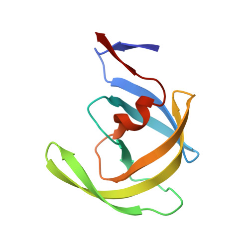
    1BV7, 1BV9, 1BWA, 1BWB

  • PubMed Abstract: 

    The long-term therapeutic benefit of HIV antiretroviral therapy is still threatened by drug-resistant variants. Mutations in the S1 subsite of the protease are the primary cause for the loss of sensitivity toward many HIV protease inhibitors, including our first-generation cyclic urea-based inhibitors DMP323 and DMP450. We now report the structures of the three active-site mutant proteases V82F, I84V, and V82F/I84V in complex with XV638 and SD146, two P2 analogues of DMP323 that are 8-fold more potent against the wild type and are able to inhibit a broad panel of drug-resistant variants [Jadhav, P. K., et al. (1997) J. Med. Chem. 40, 181-191]. The increased efficacy of XV638 and SD146 is due primarily to an increase in P2-S2 interactions: 30-40% more van der Waals contacts and two to four additional hydrogen bonds. Furthermore, because these new interactions do not perturb other subsites in the protease, it appears that the large complementary surface areas of their P2 substituents compensate for the loss of P1-S1 interactions and reduce the probability of selecting for drug-resistant variants.


  • Organizational Affiliation

    Experimental Station, DuPont Pharmaceuticals, Wilmington, Delaware 19880, USA.


Macromolecules
Find similar proteins by:  (by identity cutoff)  |  3D Structure
Entity ID: 1
MoleculeChains Sequence LengthOrganismDetailsImage
PROTEIN (HIV-1 PROTEASE)
A, B
99Human immunodeficiency virus 1Mutation(s): 2 
EC: 3.4.23.16
UniProt
Find proteins for P04585 (Human immunodeficiency virus type 1 group M subtype B (isolate HXB2))
Explore P04585 
Go to UniProtKB:  P04585
Entity Groups  
Sequence Clusters30% Identity50% Identity70% Identity90% Identity95% Identity100% Identity
UniProt GroupP04585
Sequence Annotations
Expand
  • Reference Sequence
Small Molecules
Ligands 1 Unique
IDChains Name / Formula / InChI Key2D Diagram3D Interactions
XV6
Query on XV6

Download Ideal Coordinates CCD File 
C [auth B][4R-(4ALPHA,5ALPHA,6BETA,7BETA)]-3,3'-[[TETRAHYDRO-5,6-DIHYDROXY-2-OXO-4,7-BIS(PHENYLMETHYL)-1H-1,3-DIAZEPINE-1,3(2H)-D IYL] BIS(METHYLENE)]BIS[N-2-THIAZOLYLBENZAMIDE]
C41 H38 N6 O5 S2
JDALSSGOBMTZEP-NWJWHWDBSA-N
Binding Affinity Annotations 
IDSourceBinding Affinity
XV6 Binding MOAD:  1BWA Ki: 2.5 (nM) from 1 assay(s)
PDBBind:  1BWA Ki: 2.5 (nM) from 1 assay(s)
Experimental Data & Validation

Experimental Data

  • Method: X-RAY DIFFRACTION
  • Resolution: 1.90 Å
  • R-Value Work: 0.199 
  • R-Value Observed: 0.199 
  • Space Group: P 61
Unit Cell:
Length ( Å )Angle ( ˚ )
a = 62.8α = 90
b = 62.8β = 90
c = 83.5γ = 120
Software Package:
Software NamePurpose
X-PLORrefinement

Structure Validation

View Full Validation Report


Entry History 

Deposition Data

Revision History  (Full details and data files)

  • Version 1.0: 1998-10-30
    Type: Initial release
  • Version 1.1: 2008-04-27
    Changes: Version format compliance
  • Version 1.2: 2011-07-13
    Changes: Version format compliance
  • Version 1.3: 2017-11-29
    Changes: Derived calculations
  • Version 1.4: 2021-11-03
    Changes: Database references, Derived calculations, Structure summary
  • Version 1.5: 2024-02-07
    Changes: Data collection