Three-dimensional solution structure of the HIV-1 protease complexed with DMP323, a novel cyclic urea-type inhibitor, determined by nuclear magnetic resonance spectroscopy.
Yamazaki, T., Hinck, A.P., Wang, Y.X., Nicholson, L.K., Torchia, D.A., Wingfield, P., Stahl, S.J., Kaufman, J.D., Chang, C.H., Domaille, P.J., Lam, P.Y.(1996) Protein Sci 5: 495-506
- PubMed: 8868486
- DOI: https://doi.org/10.1002/pro.5560050311
- Primary Citation of Related Structures:
1BVE, 1BVG - PubMed Abstract:
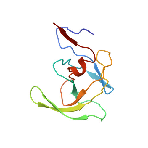
The three-dimensional solution structure of the HIV-1 protease homodimer, MW 22.2 kDa, complexed to a potent, cyclic urea-based inhibitor, DMP323, is reported. This is the first solution structure of an HIV protease/inhibitor complex that has been elucidated. Multidimensional heteronuclear NMR spectra were used to assemble more than 4,200 distance and angle constraints. Using the constraints, together with a hybrid distance geometry/simulated annealing protocol, an ensemble of 28 NMR structures was calculated having no distance or angle violations greater than 0.3 A or 5 degrees, respectively. Neglecting residues in disordered loops, the RMS deviation (RMSD) for backbone atoms in the family of structures was 0.60 A relative to the average structure. The individual NMR structures had excellent covalent geometry and stereochemistry, as did the restrained minimized average structure. The latter structure is similar to the 1.8-A X-ray structure of the protease/DMP323 complex (Chang CH et al., 1995, Protein Science, submitted); the pairwise backbone RMSD calculated for the two structures is 1.22 A. As expected, the mismatch between the structures is greatest in the loops that are disordered and/or flexible. The flexibility of residues 37-42 and 50-51 may be important in facilitating substrate binding and product release, because these residues make up the respective hinges and tips of the protease flaps. Flexibility of residues 4-8 may play a role in protease regulation by facilitating autolysis.
Organizational Affiliation:
Molecular Structural Biology Unit, NIDR, Bethesda, Maryland 20892, USA.