1BL0

MULTIPLE ANTIBIOTIC RESISTANCE PROTEIN (MARA)/DNA COMPLEX


Experimental Data Snapshot

  • Method: X-RAY DIFFRACTION
  • Resolution: 2.30 Å
  • R-Value Free: 0.303 
  • R-Value Work: 0.225 
  • R-Value Observed: 0.225 

wwPDB Validation   3D Report Full Report


This is version 1.3 of the entry. See complete history


Literature

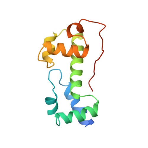
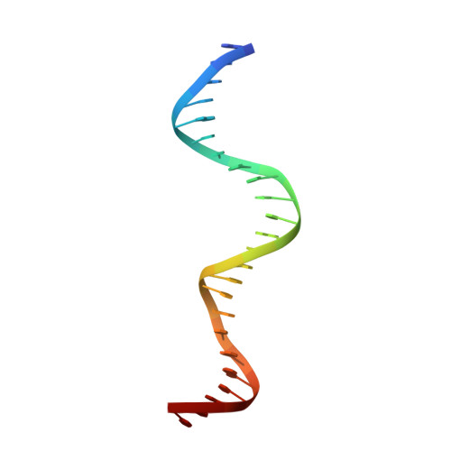
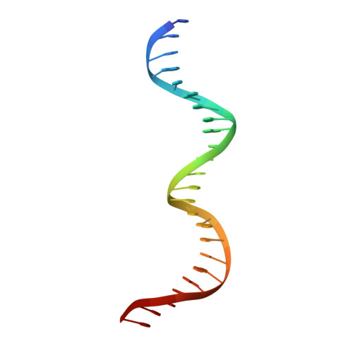
A novel DNA-binding motif in MarA: the first structure for an AraC family transcriptional activator.

Rhee, S.Martin, R.G.Rosner, J.L.Davies, D.R.

(1998) Proc Natl Acad Sci U S A 95: 10413-10418

  • DOI: https://doi.org/10.1073/pnas.95.18.10413
  • Primary Citation of Related Structures:  
    1BL0

  • PubMed Abstract: 

    A crystal structure for a member of the AraC prokaryotic transcriptional activator family, MarA, in complex with its cognate DNA-binding site is described. MarA consists of two similar subdomains, each containing a helix-turn-helix DNA-binding motif. The two recognition helices of the motifs are inserted into adjacent major groove segments on the same face of the DNA but are separated by only 27 A thereby bending the DNA by approximately 35 degrees. Extensive interactions between the recognition helices and the DNA major groove provide the sequence specificity.


  • Organizational Affiliation

    Laboratory of Molecular Biology, National Institutes of Diabetes, Digestive and Kidney Diseases, National Institutes of Health, Bethesda, MD 20892-0560, USA.


Macromolecules

Find similar proteins by:  (by identity cutoff)  |  3D Structure
Entity ID: 3
MoleculeChains Sequence LengthOrganismDetailsImage
PROTEIN (MULTIPLE ANTIBIOTIC RESISTANCE PROTEIN)C [auth A]129Escherichia coliMutation(s): 0 
Gene Names: MARA
UniProt
Find proteins for P0ACH5 (Escherichia coli (strain K12))
Explore P0ACH5 
Go to UniProtKB:  P0ACH5
Entity Groups  
Sequence Clusters30% Identity50% Identity70% Identity90% Identity95% Identity100% Identity
UniProt GroupP0ACH5
Sequence Annotations
Expand
  • Reference Sequence

Find similar nucleic acids by:  Sequence   |   3D Structure  

Entity ID: 1
MoleculeChains LengthOrganismImage
DNA (5'-D(*GP*GP*GP*GP*AP*TP*TP*TP*AP*GP*CP*AP*AP*AP*AP*CP*GP*TP*GP*GP*CP*AP* TP*C)-3')A [auth B]24N/A
Sequence Annotations
Expand
  • Reference Sequence

Find similar nucleic acids by:  Sequence   |   3D Structure  

Entity ID: 2
MoleculeChains LengthOrganismImage
DNA (5'-D(*CP*CP*GP*AP*TP*GP*CP*CP*AP*CP*GP*TP*TP*TP*TP*GP*CP*TP*AP*AP*AP*TP* CP*C)-3')B [auth C]24N/A
Sequence Annotations
Expand
  • Reference Sequence
Experimental Data & Validation

Experimental Data

  • Method: X-RAY DIFFRACTION
  • Resolution: 2.30 Å
  • R-Value Free: 0.303 
  • R-Value Work: 0.225 
  • R-Value Observed: 0.225 
  • Space Group: P 41 21 2
Unit Cell:
Length ( Å )Angle ( ˚ )
a = 47.1α = 90
b = 47.1β = 90
c = 298.2γ = 90
Software Package:
Software NamePurpose
MLPHAREphasing
X-PLORrefinement
DENZOdata reduction
SCALEPACKdata scaling

Structure Validation

View Full Validation Report


Entry History 

Deposition Data

Revision History  (Full details and data files)

  • Version 1.0: 1998-09-02
    Type: Initial release
  • Version 1.1: 2008-04-27
    Changes: Version format compliance
  • Version 1.2: 2011-07-13
    Changes: Version format compliance
  • Version 1.3: 2024-02-07
    Changes: Data collection, Database references