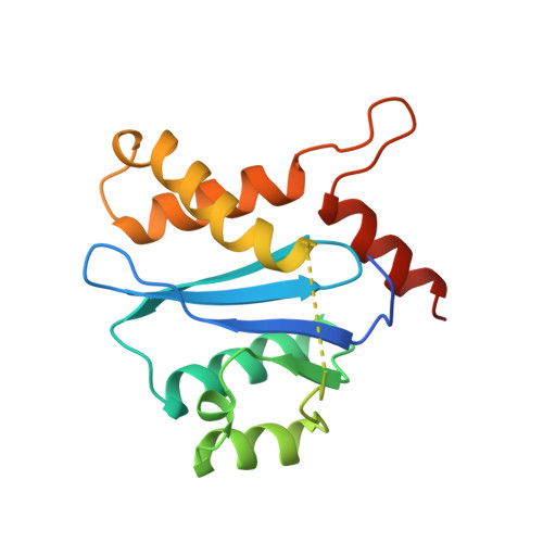
Three new structures of the core domain of HIV-1 integrase: an active site that binds magnesium.
Goldgur, Y., Dyda, F., Hickman, A.B., Jenkins, T.M., Craigie, R., Davies, D.R.(1998) Proc Natl Acad Sci U S A 95: 9150-9154
- PubMed: 9689049
- DOI: https://doi.org/10.1073/pnas.95.16.9150
- Primary Citation of Related Structures:
1BIS, 1BIU, 1BIZ - PubMed Abstract:
HIV-1 integrase is an essential enzyme in the life cycle of the virus, responsible for catalyzing the insertion of the viral genome into the host cell chromosome; it provides an attractive target for antiviral drug design. The previously reported crystal structure of the HIV-1 integrase core domain revealed that this domain belongs to the superfamily of polynucleotidyltransferases. However, the position of the conserved catalytic carboxylic acids differed from those observed in other enzymes of the class, and attempts to crystallize in the presence of the cofactor, Mg2+, were unsuccessful. We report here three additional crystal structures of the core domain of HIV-1 integrase mutants, crystallized in the presence and absence of cacodylate, as well as complexed with Mg2+. These three crystal forms, containing between them seven independent core domain structures, demonstrate the unambiguous extension of the previously disordered helix alpha4 toward the amino terminus from residue M154 and show that the catalytic E152 points in the general direction of the two catalytic aspartates, D64 and D116. In the vicinity of the active site, the structure of the protein in the absence of cacodylate exhibits significant deviations from the previously reported structures. These differences can be attributed to the modification of C65 and C130 by cacodylate, which was an essential component of the original crystallization mixture. We also demonstrate that in the absence of cacodylate this protein will bind to Mg2+, and could provide a satisfactory platform for binding of inhibitors.
Organizational Affiliation:
Laboratory of Molecular Biology, National Institute of Diabetes and Digestive and Kidney Diseases, National Institutes of Health, Bethesda, MD 20892, USA.