How a protein prepares for B12 binding: structure and dynamics of the B12-binding subunit of glutamate mutase from Clostridium tetanomorphum
Tollinger, M., Konrat, R., Hilbert, B.H., Marsh, E.N., Krautler, B.(1998) Structure 6: 1021-1033
- PubMed: 9739092
- DOI: https://doi.org/10.1016/s0969-2126(98)00103-8
- Primary Citation of Related Structures:
1BE1 - PubMed Abstract:
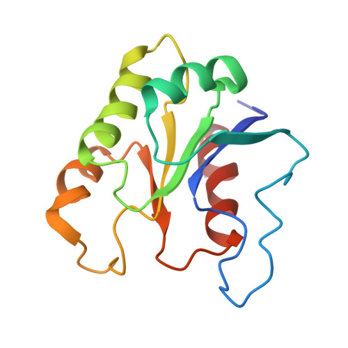
Glutamate mutase is an adenosylcobamide (coenzyme B12) dependent enzyme that catalyzes the reversible rearrangement of (2S)-glutamate to (2S,3S)-3-methylaspartate. The enzyme from Clostridium tetanomorphum comprises two subunits (of 53.7 and 14.8 kDa) and in its active form appears to be an alpha 2 beta 2 tetramer. The smaller subunit, termed MutS, has been characterized as the B12-binding component. Knowledge on the structure of a B12-binding apoenzyme does not exist. The solution structure and important dynamical aspects of MutS have been determined from a heteronuclear NMR study. The global fold of MutS in solution resembles that determined by X-ray crystallography for the B12-binding domains of Escherichia coli methionine synthase and Propionibacterium shermanii methylmalonyl CoA mutase. In these two proteins a histidine residue displaces the endogenous cobalt-coordinating ligand of the B12 cofactor. In MutS, however, the segment of the protein containing the conserved histidine residue forms part of an unstructured and mobile extended loop. A comparison of the crystal structures of two B12-binding domains, with bound B12 cofactor, and the solution structure of the apoprotein MutS has helped to clarify the mechanism of B12 binding. The major part of MutS is preorganized for B12 binding, but the B12-binding site itself is only partially formed. Upon binding B12, important elements of the binding site appear to become structured, including an alpha helix that forms one side of the cleft accommodating the nucleotide 'tail' of the cofactor.
Organizational Affiliation:
Institute of Organic Chemistry, University of Innsbruck, Austria.