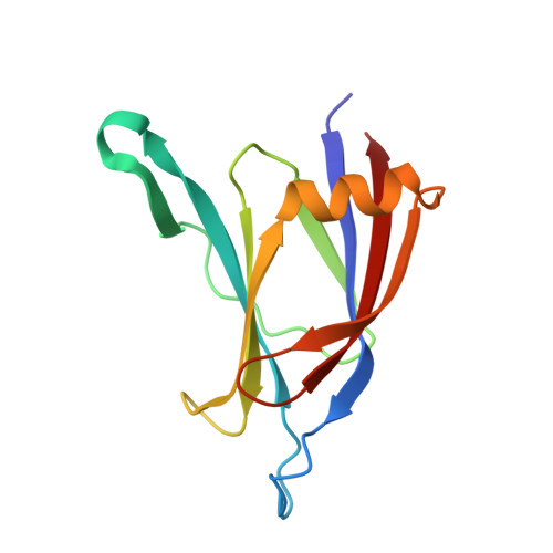
Crystal structure of the C2 domain from protein kinase C-delta.
Pappa, H., Murray-Rust, J., Dekker, L.V., Parker, P.J., McDonald, N.Q.(1998) Structure 6: 885-894
- PubMed: 9687370
- DOI: https://doi.org/10.1016/s0969-2126(98)00090-2
- Primary Citation of Related Structures:
1BDY - PubMed Abstract:
The protein kinase C (PKC) family of lipid-dependent serine/theonine kinases plays a central role in many intracellular eukaryotic signalling events. Members of the novel (delta, epsilon, eta, theta) subclass of PKC isotypes lack the Ca2+ dependence of the conventional PKC isotypes and have an N-terminal C2 domain, originally defined as V0 (variable domain zero). Biochemical data suggest that this domain serves to translocate novel PKC family members to the plasma membrane and may influence binding of PKC activators. The crystal structure of PKC-delta C2 domain indicates an unusual variant of the C2 fold. Structural elements unique to this C2 domain include a helix and a protruding beta hairpin which may contribute basic sequences to a membrane-interaction site. The invariant C2 motif, Pro-X-Trp, where X is any amino acid, forms a short crossover loop, departing radically from its conformation in other C2 structures, and contains a tyrosine phosphorylation site unique to PKC-delta. This loop and two others adopt quite different conformations from the equivalent Ca(2+)-binding loops of phospholipase C-delta and synaptotagmin I, and lack sequences necessary for Ca2+ coordination. The N-terminal sequence of Ca(2+)-independent novel PKCs defines a divergent example of a C2 structure similar to that of phospholipase C-delta. The Ca(2+)-independent regulation of novel PKCs is explained by major structural and sequence differences resulting in three non-functional Ca(2+)-binding loops. The observed structural variation and position of a tyrosine-phosphorylation site suggest the existence of distinct subclasses of C2-like domains which may have evolved distinct functional roles and mechanisms to interact with lipid membranes.
Organizational Affiliation:
Structural Biology, Imperial Cancer Research Fund, London, UK.