1B2I

KRINGLE 2 DOMAIN OF HUMAN PLASMINOGEN: NMR SOLUTION STRUCTURE OF TRANS-4-AMINOMETHYLCYCLOHEXANE-1-CARBOXYLIC ACID (AMCHA) COMPLEX


Experimental Data Snapshot

  • Method: SOLUTION NMR
  • Conformers Calculated: 150 
  • Conformers Submitted: 20 
  • Selection Criteria: TOTAL ENERGY, RAMACHANDRAN PLOT, LEAST RESTRAINT VIOLATION 

wwPDB Validation   3D Report Full Report


This is version 1.5 of the entry. See complete history


Literature

Solution structure and dynamics of the plasminogen kringle 2-AMCHA complex: 3(1)-helix in homologous domains.

Marti, D.N.Schaller, J.Llinas, M.

(1999) Biochemistry 38: 15741-15755

  • DOI: https://doi.org/10.1021/bi9917378
  • Primary Citation of Related Structures:  
    1B2I

  • PubMed Abstract: 

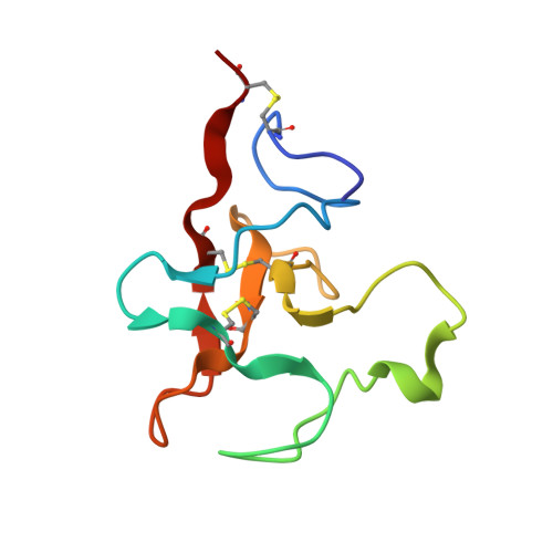
    The kringle 2 (K2) module of human plasminogen (Pgn) binds L-lysine and analogous zwitterionic compounds, such as the antifibronolytic agent trans-(aminomethyl)cyclohexanecarboxylic acid (AMCHA). Far-UV CD and NMR spectra reveal little conformational change in K2 upon ligand binding. However, retarded (1)H-(2)H isotope exchange kinetics induced by AMCHA indicate stabilization of the K2 conformation by the ligand. Assessment of secondary structure content from CD spectra yields approximately 26% beta-STRAND, approximately 13% beta-TURN, approximately 15% 3(1)-HELIX, and approximately 6% 3(10)-HELIX. The NMR solution conformation of the K2 domain complexed to AMCHA has been determined [heavy atom rmsd = 0.49 +/- 0.09A (BACKBONE) AND 1.02+/- 0.08 (ALL)]. The K2 molecule has overall dimensions of approximately 34.5A times approximately 33.4A times approximately 22.7A . Analogous with the polypeptide outline of homologous domains, K2 contains three short antiparallel beta-sheets (paired strands 15-16/20-21, 24-25/48-49, and 62-64/72-74) and four defined beta-turns (residues 6-9, 16-19, 53-56, AND 67-70). Consistent with the CD analysis, albeit novel in the context of kringle folding, the NMR structure reveals an unpaired beta-strand structured by residues 30-32, a turn of 3(10)-helix compromising residues 38-41, and a 3(1)-helix for residues 21-24 and 74-79. We also identify alignable 3(1)-helices in previously reported homologous kringle structures. Rather high order parameter S(2) values (= approximately 0.85 +/- 0.04) characterize the K2 backbone dynamics. The lowest flexibility is observed for the two inner loop segments of residues 51-63 AND 63-75 (= approximately 0.86-0.87 +/- 0.03). Overhauser connectivities reveal close hydrophobic contacts of the ligand ring with side chains of Tyr(36), Trp(62), Phe(64), Trp(72), AND Leu(74). In most K2 structures, the N atom of AMCHA places itself approximately 3.9 and 4.4A from the anionic groups of Glu(57) and Asp(55), respectively, while its carboxylate group, H-bonded to the Tyr(36) side chain OH(eta), ion-pairs the Arg(71) guanidinium group. Consistent with the preference of K2 for binding 5-aminopentanoic acid over 6-aminohexanoic acid, the positions of the ionic centers within the K2 binding site approach each other approximately 1A closer relative to what is observed in lysine binding sites of homologous Pgn modules.


  • Organizational Affiliation

    Department of Chemistry, Carnegie Mellon University, Pittsburgh, Pennsylvania 15213, and Department of Chemistry and Biochemistry, University of Bern, CH-3012 Bern, Switzerland.


Macromolecules
Find similar proteins by:  (by identity cutoff)  |  3D Structure
Entity ID: 1
MoleculeChains Sequence LengthOrganismDetailsImage
PROTEIN (PLASMINOGEN)83Homo sapiensMutation(s): 3 
EC: 3.4.21.7
UniProt & NIH Common Fund Data Resources
Find proteins for P00747 (Homo sapiens)
Explore P00747 
Go to UniProtKB:  P00747
PHAROS:  P00747
GTEx:  ENSG00000122194 
Entity Groups  
Sequence Clusters30% Identity50% Identity70% Identity90% Identity95% Identity100% Identity
UniProt GroupP00747
Sequence Annotations
Expand
  • Reference Sequence
Small Molecules
Ligands 1 Unique
IDChains Name / Formula / InChI Key2D Diagram3D Interactions
AMH
Query on AMH

Download Ideal Coordinates CCD File 
B [auth A]TRANS-4-AMINOMETHYLCYCLOHEXANE-1-CARBOXYLIC ACID
C8 H15 N O2
GYDJEQRTZSCIOI-LJGSYFOKSA-N
Binding Affinity Annotations 
IDSourceBinding Affinity
AMH PDBBind:  1B2I IC50: 3100 (nM) from 1 assay(s)
Experimental Data & Validation

Experimental Data

  • Method: SOLUTION NMR
  • Conformers Calculated: 150 
  • Conformers Submitted: 20 
  • Selection Criteria: TOTAL ENERGY, RAMACHANDRAN PLOT, LEAST RESTRAINT VIOLATION 

Structure Validation

View Full Validation Report


Entry History 

Deposition Data

Revision History  (Full details and data files)

  • Version 1.0: 1999-11-19
    Type: Initial release
  • Version 1.1: 2008-04-26
    Changes: Version format compliance
  • Version 1.2: 2011-07-13
    Changes: Version format compliance
  • Version 1.3: 2017-11-29
    Changes: Derived calculations
  • Version 1.4: 2021-11-03
    Changes: Database references, Derived calculations
  • Version 1.5: 2023-12-27
    Changes: Data collection