1A7F

INSULIN MUTANT B16 GLU, B24 GLY, DES-B30, NMR, 20 STRUCTURES


Experimental Data Snapshot

  • Method: SOLUTION NMR
  • Conformers Calculated: 50 
  • Conformers Submitted: 20 
  • Selection Criteria: LEAST TOTAL ENERGY (TARGET FUNCTION) 

wwPDB Validation   3D Report Full Report


This is version 1.3 of the entry. See complete history


Literature

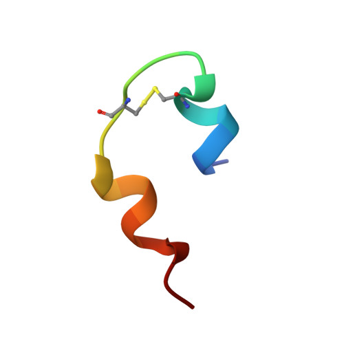
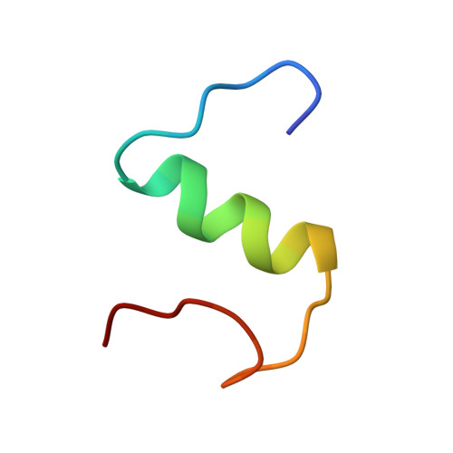
A structural switch in a mutant insulin exposes key residues for receptor binding.

Ludvigsen, S.Olsen, H.B.Kaarsholm, N.C.

(1998) J Mol Biol 279: 1-7

  • DOI: https://doi.org/10.1006/jmbi.1998.1801
  • Primary Citation of Related Structures:  
    1A7F

  • PubMed Abstract: 

    Despite years of effort to clarify the structural basis of insulin receptor binding no clear consensus has emerged. It is generally believed that insulin receptor binding is accompanied by some degree of conformational change in the carboxy-terminal of the insulin B-chain. In particular, while most substitutions for PheB24 lead to inactive species, glycine or D-amino acids are well tolerated in this position. Here we assess the conformation change by solving the solution structure of the biologically active (GluB16, GlyB24, desB30)-insulin mutant. The structure in aqueous solution at pH 8 reveals a subtle, albeit well-defined rearrangement of the C-terminal decapeptide involving a perturbation of the B20-23 turn, which allows the PheB25 residue to occupy the position normally taken up by PheB24 in native insulin. The new protein surface exposed rationalizes the receptor binding properties of a series of insulin analogs. We suggest that the structural switch is forced by the structure of the underlying core of species invariant residues and that an analogous rearrangement of the C-terminal of the B-chain occurs in native insulin on binding to its receptor.


  • Organizational Affiliation

    Novo Nordisk A/S, Bagsvaerd, Denmark.


Macromolecules

Find similar proteins by:  Sequence   |   3D Structure  

Entity ID: 1
MoleculeChains Sequence LengthOrganismDetailsImage
INSULIN21Homo sapiensMutation(s): 0 
UniProt & NIH Common Fund Data Resources
Find proteins for P01308 (Homo sapiens)
Explore P01308 
Go to UniProtKB:  P01308
PHAROS:  P01308
GTEx:  ENSG00000254647 
Entity Groups  
Sequence Clusters30% Identity50% Identity70% Identity90% Identity95% Identity100% Identity
UniProt GroupP01308
Sequence Annotations
Expand
  • Reference Sequence
Find similar proteins by:  (by identity cutoff)  |  3D Structure
Entity ID: 2
MoleculeChains Sequence LengthOrganismDetailsImage
INSULIN29Homo sapiensMutation(s): 2 
UniProt & NIH Common Fund Data Resources
Find proteins for P01308 (Homo sapiens)
Explore P01308 
Go to UniProtKB:  P01308
PHAROS:  P01308
GTEx:  ENSG00000254647 
Entity Groups  
Sequence Clusters30% Identity50% Identity70% Identity90% Identity95% Identity100% Identity
UniProt GroupP01308
Sequence Annotations
Expand
  • Reference Sequence
Experimental Data & Validation

Experimental Data

  • Method: SOLUTION NMR
  • Conformers Calculated: 50 
  • Conformers Submitted: 20 
  • Selection Criteria: LEAST TOTAL ENERGY (TARGET FUNCTION) 

Structure Validation

View Full Validation Report


Entry History 

Deposition Data

Revision History  (Full details and data files)

  • Version 1.0: 1998-07-15
    Type: Initial release
  • Version 1.1: 2008-03-24
    Changes: Version format compliance
  • Version 1.2: 2011-07-13
    Changes: Version format compliance
  • Version 1.3: 2021-11-03
    Changes: Data collection, Database references, Derived calculations, Other