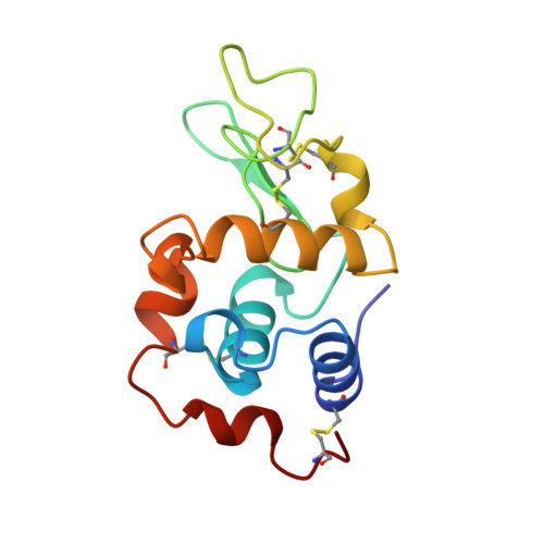
High-resolution structure (1.33 A) of a HEW lysozyme tetragonal crystal grown in the APCF apparatus. Data and structural comparison with a crystal grown under microgravity from SpaceHab-01 mission.
Vaney, M.C., Maignan, S., Ries-Kautt, M., Ducriux, A.(1996) Acta Crystallogr D Biol Crystallogr 52: 505-517
- PubMed: 15299672
- DOI: https://doi.org/10.1107/S090744499501674X
- Primary Citation of Related Structures:
193L, 194L - PubMed Abstract:
Crystals of tetragonal hen egg-white lysozyme were grown using Advanced Protein Crystallization Facility (APCF) apparatus under a microgravity environment (SpaceHab-01 mission) and ground control conditions. Crystals were grown from NaCl as a crystallizing agent at pH 4.3. The X-ray diffraction patterns of the best diffracting ground- and space-grown crystals were recorded using synchrotron radiation and an image plate on the W32 beamline at LURE. Both ground- and space-grown crystals showed nearly equivalent maximum resolution of 1.3-1.4 A. Refinements were carried out with the program X-PLOR with final R values of 18.45 and 18.27% for structures from ground- and space- grown crystals, respectively. The two structures are nearly identical with the root-mean-square difference on all protein atoms being 0.13 A. Some residues of the two refined structures show multiple alternative conformations. Two ions were localized into the electron-density maps of the two structures: one chloride ion at the interface between two symmetry-related molecules and one sodium ion stabilizing the loop Ser60-Leu75. The sodium ion is surrounded by six ligands which form a bipyramid around it at distances of 2.2-2.6 A.
Organizational Affiliation:
Laboratoire de Biologie Structurale, Bâtimet 34, UMR 9920, CNRS, Université Paris-Sud, Gif sur Yvette, France.