12E8

2E8 FAB FRAGMENT


Experimental Data Snapshot

  • Method: X-RAY DIFFRACTION
  • Resolution: 1.90 Å
  • R-Value Free: 0.271 
  • R-Value Work: 0.221 
  • R-Value Observed: 0.221 

wwPDB Validation   3D Report Full Report


This is version 1.3 of the entry. See complete history


Literature

Structure of a monoclonal 2E8 Fab antibody fragment specific for the low-density lipoprotein-receptor binding region of apolipoprotein E refined at 1.9 A.

Trakhanov, S.Parkin, S.Raffai, R.Milne, R.Newhouse, Y.M.Weisgraber, K.H.Rupp, B.

(1999) Acta Crystallogr D Biol Crystallogr null: 122-128

  • DOI: https://doi.org/10.1107/S090744499800938X
  • Primary Citation of Related Structures:  
    12E8

  • PubMed Abstract: 

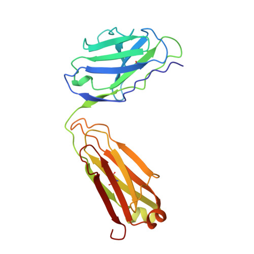
    The crystal structure of the Fab fragment of 2E8, the monoclonal IgG1,kappa antibody specific for the low-density lipoprotein (LDL) receptor-binding region of apolipoprotein E (apoE), has been solved by molecular replacement and refined at 1.9 A resolution (PDB entry 12E8). Two 2E8 Fab molecules in the asymmetric unit are related by noncrystallographic symmetry and are hydrogen bonded through a beta-sheet-like intermolecular contact between the heavy-chain complementarity-determining regions 3 (CDRH3) of each molecule. The structure has been refined to an R value of 0.22 (Rfree = 0.27). The initially ill-defined heavy-chain constant domain (CH1) of 2E8 has been retraced with the aid of automatic refinement, confirming the beta-sheet tracing independently of any starting models. As a resolution better than 2 A is not common for Fab fragments, this model represents a well defined Fab structure and should prove useful in MR solution of other Fab fragments. Furthermore, in the absence of an LDL-receptor structure, the homology of the 2E8 CDRH2 to the ligand-binding domain of the LDL receptor has been exploited to model the apoE-LDL-receptor interaction.


  • Organizational Affiliation

    Gladstone Institute of Cardiovascular Disease, University of California, San Francisco, CA 94141, USA.


Macromolecules
Find similar proteins by:  (by identity cutoff)  |  3D Structure
Entity ID: 1
MoleculeChains Sequence LengthOrganismDetailsImage
IGG1-KAPPA 2E8 FAB (LIGHT CHAIN)A [auth L],
C [auth M]
214Mus musculusMutation(s): 0 
Entity Groups  
Sequence Clusters30% Identity50% Identity70% Identity90% Identity95% Identity100% Identity
Sequence Annotations
Expand
  • Reference Sequence
Find similar proteins by:  (by identity cutoff)  |  3D Structure
Entity ID: 2
MoleculeChains Sequence LengthOrganismDetailsImage
IGG1-KAPPA 2E8 FAB (HEAVY CHAIN)B [auth H],
D [auth P]
221Mus musculusMutation(s): 0 
Entity Groups  
Sequence Clusters30% Identity50% Identity70% Identity90% Identity95% Identity100% Identity
Sequence Annotations
Expand
  • Reference Sequence
Experimental Data & Validation

Experimental Data

  • Method: X-RAY DIFFRACTION
  • Resolution: 1.90 Å
  • R-Value Free: 0.271 
  • R-Value Work: 0.221 
  • R-Value Observed: 0.221 
  • Space Group: P 1 21 1
Unit Cell:
Length ( Å )Angle ( ˚ )
a = 66.56α = 90
b = 65.57β = 97.04
c = 103.22γ = 90
Software Package:
Software NamePurpose
UCSDdata collection
UCSDdata reduction
X-PLORmodel building
X-PLORrefinement
UCSDdata scaling
X-PLORphasing

Structure Validation

View Full Validation Report


Entry History 

Deposition Data

Revision History  (Full details and data files)

  • Version 1.0: 1998-08-05
    Type: Initial release
  • Version 1.1: 2008-03-24
    Changes: Version format compliance
  • Version 1.2: 2011-07-13
    Changes: Derived calculations, Version format compliance
  • Version 1.3: 2023-08-02
    Changes: Database references, Other, Refinement description