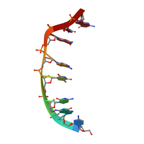
Solution structure of a DNA complex with the fluorescent bis-intercalator TOTO determined by NMR spectroscopy.
Spielmann, H.P., Wemmer, D.E., Jacobsen, J.P.(1995) Biochemistry 34: 8542-8553
- PubMed: 7612596
- DOI: https://doi.org/10.1021/bi00027a004
- Primary Citation of Related Structures:
108D - PubMed Abstract:
We have used two-dimensional 1H NMR spectroscopy to determine the solution structure of the DNA oligonucleotide d(5'-CGCTAGCG-3')2 complexed with the bis-intercalating dye 1,1'-(4,4,8,8-tetramethyl-4,8-diazaundecamethylene)bis[4-(3-methyl -2,3- dihydrobenzo-1,3-thiazolyl-2-methylidene)qui nolinium] tetraiodide (TOTO). The determination of the structure was based on total relaxation matrix analysis of the NOESY cross-peak intensities using the program MARDIGRAS. Improved procedures to consider the experimental "noise" in NOESY spectra during these calculations have been employed. The NOE-derived distance restraints were applied in restrained molecular dynamics calculations. Twenty final structures each were generated for the TOTO complex from both A-form and B-form dsDNA starting structures. The root-mean-square (rms) deviation of the coordinates for the 40 structures of the complex was 1.45 A. The local DNA structure is distorted in the complex. The helix is unwound by 60 degrees and has an overall helical repeat of 12 base pairs, caused by bis-intercalation of TOTO. The poly(propylenamine) linker chain is located in the minor groove of dsDNA. Calculations indicate that the benzothiazole ring system is twisted relative to the quinoline in the uncomplexed TOTO molecule. The site selectivity of TOTO for the CTAG-CTAG site is explained by its ability to adapt to the base pair propeller twist of dsDNA to optimize stacking and the hydrophobic interaction between the thymidine methyl group and the benzothiazole ring. There is a 3000-fold fluorescence enhancement upon binding of TOTO to dsDNA. Rotation about the cyanine methine bonds is possible in free TOTO, allowing relaxation nonradiatively. When bound to dsDNA, the benzothiazole ring and the quinolinium ring are clamped by the nucleobases preventing this rotation, and the chromophore loses excitation energy by fluorescence instead.
Organizational Affiliation:
Department of Chemistry, Odense University, Denmark.