7KJB

Crystal structure of the EphA2 S897E/S901E mutant intracellular KD-SAM domains


Experimental Data Snapshot

  • Method: X-RAY DIFFRACTION
  • Resolution: 2.80 Å
  • R-Value Free: 0.246 
  • R-Value Work: 0.198 
  • R-Value Observed: 0.200 

wwPDB Validation   3D Report Full Report


This is version 1.2 of the entry. See complete history


Literature

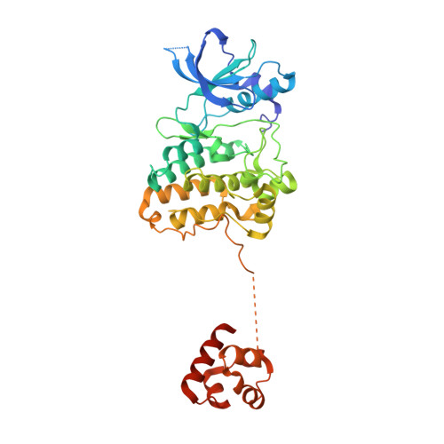
Regulation of the EphA2 receptor intracellular region by phosphomimetic negative charges in the kinase-SAM linker.

Lechtenberg, B.C.Gehring, M.P.Light, T.P.Horne, C.R.Matsumoto, M.W.Hristova, K.Pasquale, E.B.

(2021) Nat Commun 12: 7047-7047

  • DOI: https://doi.org/10.1038/s41467-021-27343-z
  • Primary Citation of Related Structures:  
    7KJA, 7KJB, 7KJC

  • PubMed Abstract: 

    Eph receptor tyrosine kinases play a key role in cell-cell communication. Lack of structural information on the entire multi-domain intracellular region of any Eph receptor has hindered understanding of their signaling mechanisms. Here, we use integrative structural biology to investigate the structure and dynamics of the EphA2 intracellular region. EphA2 promotes cancer malignancy through a poorly understood non-canonical form of signaling involving serine/threonine phosphorylation of the linker connecting its kinase and SAM domains. We show that accumulation of multiple linker negative charges, mimicking phosphorylation, induces cooperative changes in the EphA2 intracellular region from more closed to more extended conformations and perturbs the EphA2 juxtamembrane segment and kinase domain. In cells, linker negative charges promote EphA2 oligomerization. We also identify multiple kinases catalyzing linker phosphorylation. Our findings suggest multiple effects of linker phosphorylation on EphA2 signaling and imply that coordination of different kinases is necessary to promote EphA2 non-canonical signaling.


  • Organizational Affiliation

    Cancer Center, Sanford Burnham Prebys Medical Discovery Institute, La Jolla, CA, 92037, USA. lechtenberg.b@wehi.edu.au.


Macromolecules
Find similar proteins by:  (by identity cutoff)  |  3D Structure
Entity ID: 1
MoleculeChains Sequence LengthOrganismDetailsImage
Ephrin type-A receptor 2390Homo sapiensMutation(s): 2 
Gene Names: EPHA2ECK
EC: 2.7.10.1
UniProt & NIH Common Fund Data Resources
Find proteins for P29317 (Homo sapiens)
Explore P29317 
Go to UniProtKB:  P29317
PHAROS:  P29317
GTEx:  ENSG00000142627 
Entity Groups  
Sequence Clusters30% Identity50% Identity70% Identity90% Identity95% Identity100% Identity
UniProt GroupP29317
Sequence Annotations
Expand
  • Reference Sequence
Small Molecules
Ligands 1 Unique
IDChains Name / Formula / InChI Key2D Diagram3D Interactions
CS
Query on CS

Download Ideal Coordinates CCD File 
B [auth A]CESIUM ION
Cs
NCMHKCKGHRPLCM-UHFFFAOYSA-N
Experimental Data & Validation

Experimental Data

  • Method: X-RAY DIFFRACTION
  • Resolution: 2.80 Å
  • R-Value Free: 0.246 
  • R-Value Work: 0.198 
  • R-Value Observed: 0.200 
  • Space Group: P 32 2 1
Unit Cell:
Length ( Å )Angle ( ˚ )
a = 94.456α = 90
b = 94.456β = 90
c = 99.646γ = 120
Software Package:
Software NamePurpose
CrystalCleardata collection
XDSdata reduction
Aimlessdata scaling
PHASERphasing
PHENIXrefinement

Structure Validation

View Full Validation Report


Entry History & Funding Information

Deposition Data


Funding OrganizationLocationGrant Number
National Institutes of Health/National Institute of General Medical Sciences (NIH/NIGMS)United StatesGM131374

Revision History  (Full details and data files)

  • Version 1.0: 2021-02-03
    Type: Initial release
  • Version 1.1: 2022-01-12
    Changes: Database references
  • Version 1.2: 2023-10-18
    Changes: Data collection, Refinement description