7DH6

Crystal structure of PLRG1


Experimental Data Snapshot

  • Method: X-RAY DIFFRACTION
  • Resolution: 2.58 Å
  • R-Value Free: 0.276 
  • R-Value Work: 0.226 
  • R-Value Observed: 0.229 

wwPDB Validation   3D Report Full Report


This is version 1.2 of the entry. See complete history


Literature

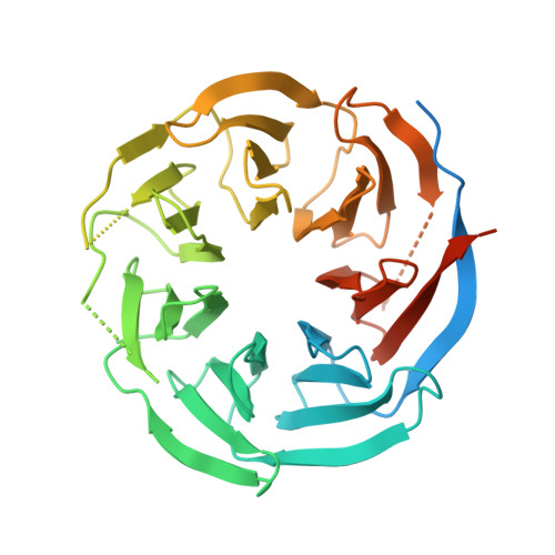
Crystal structure of the WD40 domain of human PLRG1.

Wang, X.Li, Y.Dai, H.Xu, C.

(2021) Biochem Biophys Res Commun 534: 474-477

  • DOI: https://doi.org/10.1016/j.bbrc.2020.11.057
  • Primary Citation of Related Structures:  
    7DH6

  • PubMed Abstract: 

    PLRG1 is a evolutionarily conserved protein in spliceosome and plays an important role in maintaining the integral part of the splicoeosme and its proper splicing. Here we solved the high resolution crystal structure of the WD40 domain of human PLRG1 by crystallography and compared our crystal structure with the cryo-EM structure of PLRG1 bound with other splicing factors. We found that two loops of the WD40 domain become resolved upon binding to the proteins within the spliceosome. Thus our work characterize the dynamic property of PLRG1 during the spliceosome assembly by presenting its apo structure.


  • Organizational Affiliation

    MOE Key Laboratory for Membraneless Organelles and Cellular Dynamics, Hefei National Laboratory for Physical Sciences at the Microscale and School of Life Sciences, University of Science and Technology of China, Hefei, 230027, China.


Macromolecules
Find similar proteins by:  (by identity cutoff)  |  3D Structure
Entity ID: 1
MoleculeChains Sequence LengthOrganismDetailsImage
Pleiotropic regulator 1
A, B, C, D
375Homo sapiensMutation(s): 0 
Gene Names: PLRG1
UniProt & NIH Common Fund Data Resources
Find proteins for O43660 (Homo sapiens)
Explore O43660 
Go to UniProtKB:  O43660
PHAROS:  O43660
GTEx:  ENSG00000171566 
Entity Groups  
Sequence Clusters30% Identity50% Identity70% Identity90% Identity95% Identity100% Identity
UniProt GroupO43660
Sequence Annotations
Expand
  • Reference Sequence
Experimental Data & Validation

Experimental Data

  • Method: X-RAY DIFFRACTION
  • Resolution: 2.58 Å
  • R-Value Free: 0.276 
  • R-Value Work: 0.226 
  • R-Value Observed: 0.229 
  • Space Group: P 1 21 1
Unit Cell:
Length ( Å )Angle ( ˚ )
a = 68.005α = 90
b = 82.124β = 99.44
c = 99.388γ = 90
Software Package:
Software NamePurpose
PHENIXrefinement
PDB_EXTRACTdata extraction
HKL-2000data reduction
HKL-2000data scaling
PHENIXphasing

Structure Validation

View Full Validation Report


Entry History & Funding Information

Deposition Data


Funding OrganizationLocationGrant Number
National Natural Science Foundation of China (NSFC)China--

Revision History  (Full details and data files)

  • Version 1.0: 2020-12-02
    Type: Initial release
  • Version 1.1: 2021-06-16
    Changes: Database references
  • Version 1.2: 2023-11-29
    Changes: Data collection, Database references, Refinement description