7DC8

Crystal structure of Switch Ab Fab and hIL6R in complex with ATP


Experimental Data Snapshot

  • Method: X-RAY DIFFRACTION
  • Resolution: 2.76 Å
  • R-Value Free: 0.278 
  • R-Value Work: 0.207 
  • R-Value Observed: 0.211 

wwPDB Validation   3D Report Full Report


Ligand Structure Quality Assessment 


This is version 1.1 of the entry. See complete history


Literature

Exploitation of Elevated Extracellular ATP to Specifically Direct Antibody to Tumor Microenvironment.

Mimoto, F.Tatsumi, K.Shimizu, S.Kadono, S.Haraya, K.Nagayasu, M.Suzuki, Y.Fujii, E.Kamimura, M.Hayasaka, A.Kawauchi, H.Ohara, K.Matsushita, M.Baba, T.Susumu, H.Sakashita, T.Muraoka, T.Aso, K.Katada, H.Tanaka, E.Nakagawa, K.Hasegawa, M.Ayabe, M.Yamamoto, T.Tanba, S.Ishiguro, T.Kamikawa, T.Nambu, T.Kibayashi, T.Azuma, Y.Tomii, Y.Kato, A.Ozeki, K.Murao, N.Endo, M.Kikuta, J.Kamata-Sakurai, M.Ishii, M.Hattori, K.Igawa, T.

(2020) Cell Rep 33: 108542-108542

  • DOI: https://doi.org/10.1016/j.celrep.2020.108542
  • Primary Citation of Related Structures:  
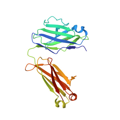
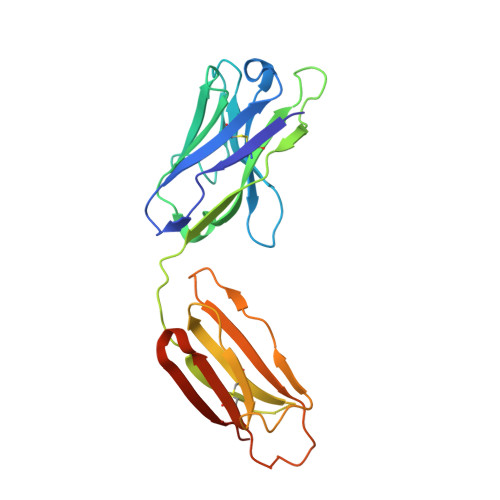
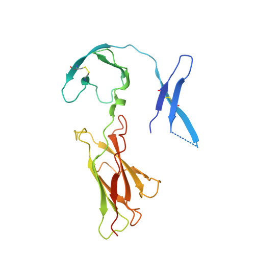
    7DC7, 7DC8

  • PubMed Abstract: 

    The extracellular adenosine triphosphate (ATP) concentration is highly elevated in the tumor microenvironment (TME) and remains tightly regulated in normal tissues. Using phage display technology, we establish a method to identify an antibody that can bind to an antigen only in the presence of ATP. Crystallography analysis reveals that ATP bound in between the antibody-antigen interface serves as a switch for antigen binding. In a transgenic mouse model overexpressing the antigen systemically, the ATP switch antibody binds to the antigen in tumors with minimal binding in normal tissues and plasma and inhibits tumor growth. Thus, we demonstrate that elevated extracellular ATP concentration can be exploited to specifically target the TME, giving therapeutic antibodies the ability to overcome on-target off-tumor toxicity.


  • Organizational Affiliation

    Chugai Pharmabody Research Pte. Ltd., 3 Biopolis Drive, #07 - 11 to 16, Synapse, 138623, Singapore. Electronic address: mimoto-f@chugai-pharmabody.com.


Macromolecules
Find similar proteins by:  (by identity cutoff)  |  3D Structure
Entity ID: 1
MoleculeChains Sequence LengthOrganismDetailsImage
Switch Ab Fab light chain
A, D
216Homo sapiensMutation(s): 0 
Entity Groups  
Sequence Clusters30% Identity50% Identity70% Identity90% Identity95% Identity100% Identity
Sequence Annotations
Expand
  • Reference Sequence
Find similar proteins by:  (by identity cutoff)  |  3D Structure
Entity ID: 2
MoleculeChains Sequence LengthOrganismDetailsImage
Switch Ab Fab heavy chain
B, E
230Homo sapiensMutation(s): 0 
Entity Groups  
Sequence Clusters30% Identity50% Identity70% Identity90% Identity95% Identity100% Identity
Sequence Annotations
Expand
  • Reference Sequence
Find similar proteins by:  (by identity cutoff)  |  3D Structure
Entity ID: 3
MoleculeChains Sequence LengthOrganismDetailsImage
Interleukin-6 receptor subunit alpha
C, F
218Homo sapiensMutation(s): 1 
Gene Names: IL6R
UniProt & NIH Common Fund Data Resources
Find proteins for P08887 (Homo sapiens)
Explore P08887 
Go to UniProtKB:  P08887
PHAROS:  P08887
GTEx:  ENSG00000160712 
Entity Groups  
Sequence Clusters30% Identity50% Identity70% Identity90% Identity95% Identity100% Identity
UniProt GroupP08887
Sequence Annotations
Expand
  • Reference Sequence
Small Molecules
Ligands 2 Unique
IDChains Name / Formula / InChI Key2D Diagram3D Interactions
ATP (Subject of Investigation/LOI)
Query on ATP

Download Ideal Coordinates CCD File 
H [auth B],
O [auth E]
ADENOSINE-5'-TRIPHOSPHATE
C10 H16 N5 O13 P3
ZKHQWZAMYRWXGA-KQYNXXCUSA-N
SO4
Query on SO4

Download Ideal Coordinates CCD File 
G [auth A]
I [auth B]
J [auth B]
K [auth C]
L [auth C]
G [auth A],
I [auth B],
J [auth B],
K [auth C],
L [auth C],
M [auth D],
N [auth D],
P [auth E],
Q [auth F],
R [auth F]
SULFATE ION
O4 S
QAOWNCQODCNURD-UHFFFAOYSA-L
Experimental Data & Validation

Experimental Data

  • Method: X-RAY DIFFRACTION
  • Resolution: 2.76 Å
  • R-Value Free: 0.278 
  • R-Value Work: 0.207 
  • R-Value Observed: 0.211 
  • Space Group: C 2 2 2
Unit Cell:
Length ( Å )Angle ( ˚ )
a = 103.043α = 90
b = 185.612β = 90
c = 152.085γ = 90
Software Package:
Software NamePurpose
PHENIXrefinement
Aimlessdata scaling
PDB_EXTRACTdata extraction
PHASERphasing
XDSdata reduction

Structure Validation

View Full Validation Report


Ligand Structure Quality Assessment 


Entry History 

Deposition Data

Revision History  (Full details and data files)

  • Version 1.0: 2021-01-13
    Type: Initial release
  • Version 1.1: 2023-11-29
    Changes: Data collection, Database references, Refinement description