


The Crystal Structure of human JMJD2A from Biortus.
Wang, F., Lv, Z., Cheng, W., Lin, D., Ju, C., Bao, X., Zhu, B.To be published.
Experimental Data Snapshot
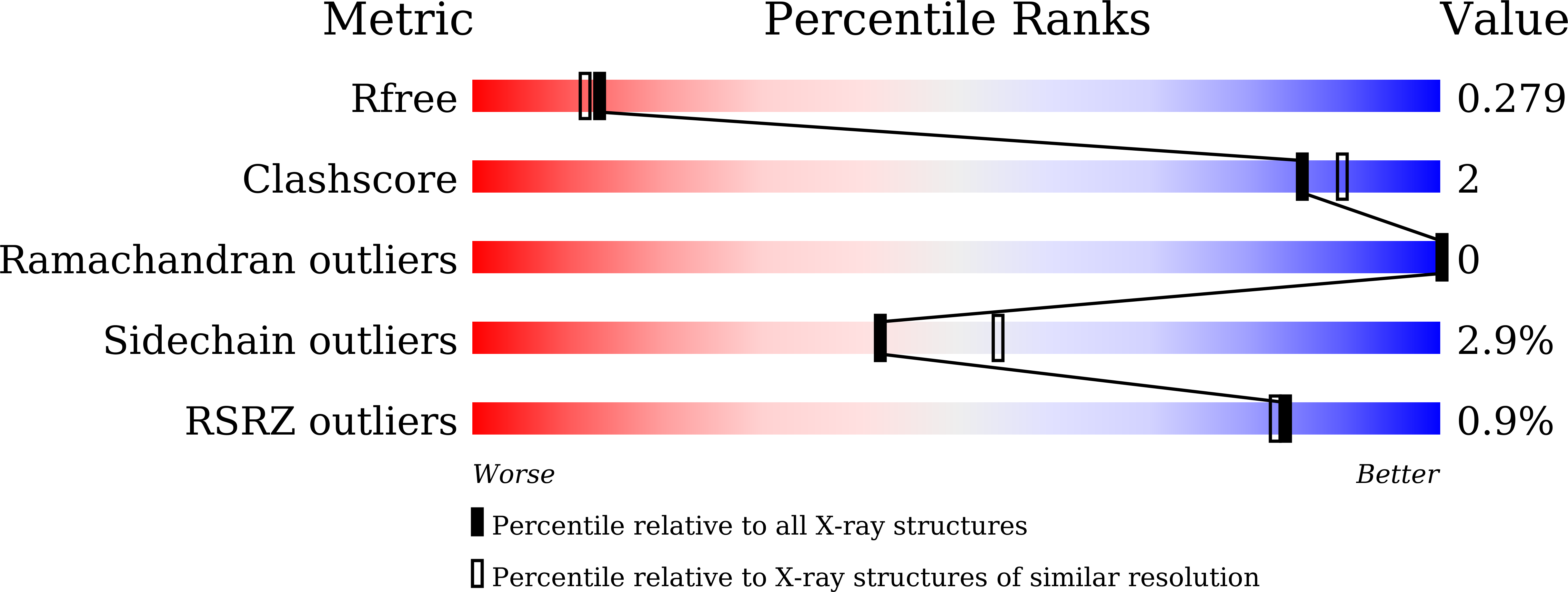
wwPDB Validation 3D Report Full Report
Entity ID: 1 | |||||
|---|---|---|---|---|---|
| Molecule | Chains | Sequence Length | Organism | Details | Image |
| Lysine-specific demethylase 4A | 131 | Homo sapiens | Mutation(s): 0 Gene Names: KDM4A, JHDM3A, JMJD2, JMJD2A, KIAA0677 EC: 1.14.11.66 (PDB Primary Data), 1.14.11.69 (PDB Primary Data) | 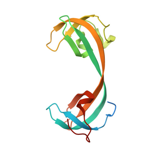 | |
UniProt & NIH Common Fund Data Resources | |||||
Find proteins for O75164 (Homo sapiens) Explore O75164 Go to UniProtKB: O75164 | |||||
PHAROS: O75164 GTEx: ENSG00000066135 | |||||
Entity Groups | |||||
| Sequence Clusters | 30% Identity50% Identity70% Identity90% Identity95% Identity100% Identity | ||||
| UniProt Group | O75164 | ||||
Sequence AnnotationsExpand | |||||
| |||||
| Ligands 1 Unique | |||||
|---|---|---|---|---|---|
| ID | Chains | Name / Formula / InChI Key | 2D Diagram | 3D Interactions | |
| SO4 Query on SO4 | B [auth A] | SULFATE ION O4 S QAOWNCQODCNURD-UHFFFAOYSA-L |  | ||
| Length ( Å ) | Angle ( ˚ ) |
|---|---|
| a = 135.11 | α = 90 |
| b = 135.11 | β = 90 |
| c = 135.11 | γ = 90 |
| Software Name | Purpose |
|---|---|
| REFMAC | refinement |
| XDS | data reduction |
| Aimless | data scaling |
| PHASER | phasing |
RCSB PDB (citation) is hosted by
RCSB PDB is a member of the

