7CO1

Crystal structure of SMAD2 in complex with wild-type CBP


Experimental Data Snapshot

  • Method: X-RAY DIFFRACTION
  • Resolution: 3.30 Å
  • R-Value Free: 0.283 
  • R-Value Work: 0.255 
  • R-Value Observed: 0.257 

wwPDB Validation   3D Report Full Report


This is version 1.2 of the entry. See complete history


Literature

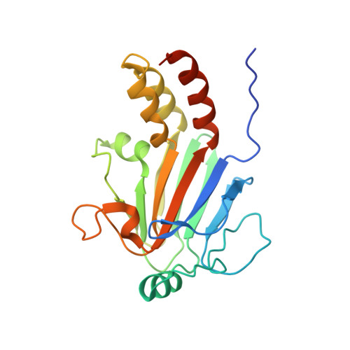
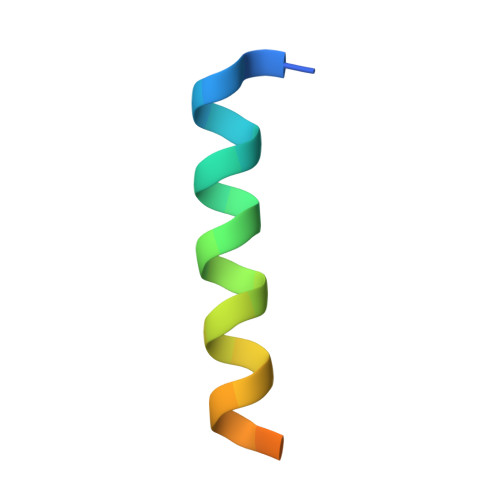
Structural basis for transcriptional coactivator recognition by SMAD2 in TGF-beta signaling.

Miyazono, K.I.Ito, T.Fukatsu, Y.Wada, H.Kurisaki, A.Tanokura, M.

(2020) Sci Signal 13

  • DOI: https://doi.org/10.1126/scisignal.abb9043
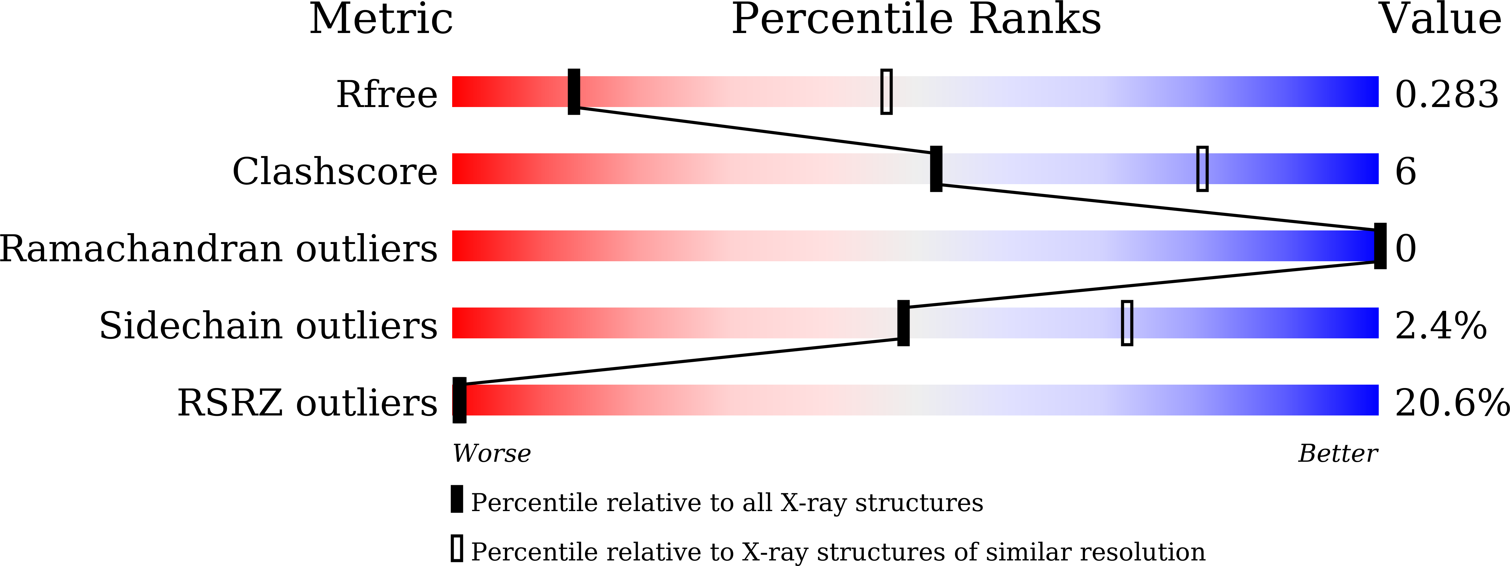
  • Primary Citation of Related Structures:  
    6M64, 7CO1

  • PubMed Abstract: 

    Transforming growth factor-β (TGF-β) proteins regulate multiple cellular functions, including cell proliferation, apoptosis, and extracellular matrix formation. The dysregulation of TGF-β signaling causes diseases such as cancer and fibrosis, and therefore, understanding the biochemical basis of TGF-β signal transduction is important for elucidating pathogenic mechanisms in these diseases. SMAD proteins are transcription factors that mediate TGF-β signaling-dependent gene expression. The transcriptional coactivator CBP directly interacts with the MH2 domains of SMAD2 to activate SMAD complex-dependent gene expression. Here, we report the structural basis for CBP recognition by SMAD2. The crystal structures of the SMAD2 MH2 domain in complex with the SMAD2-binding region of CBP showed that CBP forms an amphiphilic helix on the hydrophobic surface of SMAD2. The expression of a mutated CBP peptide that showed increased SMAD2 binding repressed SMAD2-dependent gene expression in response to TGF-β signaling in cultured cells. Disrupting the interaction between SMAD2 and CBP may therefore be a promising strategy for suppressing SMAD-dependent gene expression.


  • Organizational Affiliation

    Department of Applied Biological Chemistry, Graduate School of Agricultural and Life Sciences, The University of Tokyo, Tokyo 113-8657, Japan.


Macromolecules
Find similar proteins by:  (by identity cutoff)  |  3D Structure
Entity ID: 1
MoleculeChains Sequence LengthOrganismDetailsImage
Mothers against decapentaplegic homolog 2
A, C, E
208Homo sapiensMutation(s): 2 
Gene Names: SMAD2MADH2MADR2
UniProt & NIH Common Fund Data Resources
Find proteins for Q15796 (Homo sapiens)
Explore Q15796 
Go to UniProtKB:  Q15796
PHAROS:  Q15796
GTEx:  ENSG00000175387 
Entity Groups  
Sequence Clusters30% Identity50% Identity70% Identity90% Identity95% Identity100% Identity
UniProt GroupQ15796
Sequence Annotations
Expand
  • Reference Sequence
Find similar proteins by:  (by identity cutoff)  |  3D Structure
Entity ID: 2
MoleculeChains Sequence LengthOrganismDetailsImage
CREB-binding protein
B, D, F
28Homo sapiensMutation(s): 0 
Gene Names: CREBBPCBP
EC: 2.3.1.48 (PDB Primary Data), 2.3.1 (PDB Primary Data)
UniProt & NIH Common Fund Data Resources
Find proteins for Q92793 (Homo sapiens)
Explore Q92793 
Go to UniProtKB:  Q92793
PHAROS:  Q92793
GTEx:  ENSG00000005339 
Entity Groups  
Sequence Clusters30% Identity50% Identity70% Identity90% Identity95% Identity100% Identity
UniProt GroupQ92793
Sequence Annotations
Expand
  • Reference Sequence
Experimental Data & Validation

Experimental Data

  • Method: X-RAY DIFFRACTION
  • Resolution: 3.30 Å
  • R-Value Free: 0.283 
  • R-Value Work: 0.255 
  • R-Value Observed: 0.257 
  • Space Group: P 21 21 21
Unit Cell:
Length ( Å )Angle ( ˚ )
a = 68.922α = 90
b = 70.395β = 90
c = 163.954γ = 90
Software Package:
Software NamePurpose
Aimlessdata scaling
PHENIXrefinement
PDB_EXTRACTdata extraction
XDSdata reduction
MOLREPphasing

Structure Validation

View Full Validation Report


Entry History & Funding Information

Deposition Data


Funding OrganizationLocationGrant Number
Japan Society for the Promotion of Science (JSPS)Japan15K14708
Japan Society for the Promotion of Science (JSPS)Japan17K19581
Japan Society for the Promotion of Science (JSPS)Japan23228003
Japan Society for the Promotion of Science (JSPS)Japan20H02910

Revision History  (Full details and data files)

  • Version 1.0: 2020-11-25
    Type: Initial release
  • Version 1.1: 2020-12-30
    Changes: Database references
  • Version 1.2: 2023-11-29
    Changes: Data collection, Database references, Refinement description