7C8S

Crystal structure of DUSP22 mutant_N128A


Experimental Data Snapshot

  • Method: X-RAY DIFFRACTION
  • Resolution: 1.31 Å
  • R-Value Free: 0.197 
  • R-Value Work: 0.187 
  • R-Value Observed: 0.188 

wwPDB Validation   3D Report Full Report


Ligand Structure Quality Assessment 


This is version 1.1 of the entry. See complete history


Literature

Structural Insights into the Active Site Formation of DUSP22 in N-loop-containing Protein Tyrosine Phosphatases.

Lai, C.H.Chang, C.C.Chuang, H.C.Tan, T.H.Lyu, P.C.

(2020) Int J Mol Sci 21

  • DOI: https://doi.org/10.3390/ijms21207515
  • Primary Citation of Related Structures:  
    6L1S, 6LMY, 6LOT, 6LOU, 6LVQ, 7C8S

  • PubMed Abstract: 

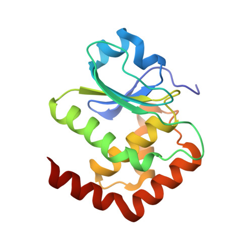
    Cysteine-based protein tyrosine phosphatases (Cys-based PTPs) perform dephosphorylation to regulate signaling pathways in cellular responses. The hydrogen bonding network in their active site plays an important conformational role and supports the phosphatase activity. Nearly half of dual-specificity phosphatases (DUSPs) use three conserved residues, including aspartate in the D-loop, serine in the P-loop, and asparagine in the N-loop, to form the hydrogen bonding network, the D-, P-, N-triloop interaction (DPN-triloop interaction). In this study, DUSP22 is used to investigate the importance of the DPN-triloop interaction in active site formation. Alanine mutations and somatic mutations of the conserved residues, D57, S93, and N128 substantially decrease catalytic efficiency ( k cat / K M ) by more than 10 2 -fold. Structural studies by NMR and crystallography reveal that each residue can perturb the three loops and induce conformational changes, indicating that the hydrogen bonding network aligns the residues in the correct positions for substrate interaction and catalysis. Studying the DPN-triloop interaction reveals the mechanism maintaining phosphatase activity in N-loop-containing PTPs and provides a foundation for further investigation of active site formation in different members of this protein class.


  • Organizational Affiliation

    Institute of Bioinformatics and Structural Biology, Department of Life Science, National Tsing Hua University, Hsinchu 30013, Taiwan.


Macromolecules
Find similar proteins by:  (by identity cutoff)  |  3D Structure
Entity ID: 1
MoleculeChains Sequence LengthOrganismDetailsImage
Dual specificity protein phosphatase 22157Homo sapiensMutation(s): 1 
Gene Names: DUSP22JSP1LMWDSP2MKPX
EC: 3.1.3.16 (PDB Primary Data), 3.1.3.48 (PDB Primary Data)
UniProt & NIH Common Fund Data Resources
Find proteins for Q9NRW4 (Homo sapiens)
Explore Q9NRW4 
Go to UniProtKB:  Q9NRW4
PHAROS:  Q9NRW4
GTEx:  ENSG00000112679 
Entity Groups  
Sequence Clusters30% Identity50% Identity70% Identity90% Identity95% Identity100% Identity
UniProt GroupQ9NRW4
Sequence Annotations
Expand
  • Reference Sequence
Small Molecules
Ligands 1 Unique
IDChains Name / Formula / InChI Key2D Diagram3D Interactions
SO4 (Subject of Investigation/LOI)
Query on SO4

Download Ideal Coordinates CCD File 
B [auth A]SULFATE ION
O4 S
QAOWNCQODCNURD-UHFFFAOYSA-L
Experimental Data & Validation

Experimental Data

  • Method: X-RAY DIFFRACTION
  • Resolution: 1.31 Å
  • R-Value Free: 0.197 
  • R-Value Work: 0.187 
  • R-Value Observed: 0.188 
  • Space Group: C 1 2 1
Unit Cell:
Length ( Å )Angle ( ˚ )
a = 89.842α = 90
b = 50.432β = 95.96
c = 40.212γ = 90
Software Package:
Software NamePurpose
HKL-2000data reduction
HKL-2000data scaling
MOLREPphasing
PHENIXrefinement
PDB_EXTRACTdata extraction

Structure Validation

View Full Validation Report


Ligand Structure Quality Assessment 


Entry History & Funding Information

Deposition Data


Funding OrganizationLocationGrant Number
Ministry of Science and Technology (MoST, Taiwan)Taiwan--

Revision History  (Full details and data files)

  • Version 1.0: 2020-10-28
    Type: Initial release
  • Version 1.1: 2023-11-29
    Changes: Data collection, Database references, Refinement description