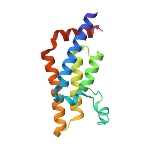
6YB4

Crystal structure of human ATAD2 bromodomain in complex with N-(4-bromo-3-(3-methylpyrrolidin-1-yl)sulfonyl)phenyl)-2-(-4-cyclopropyl-4-methyl-2,5-dioxoimidazolidin-1-yl)acetamide


Experimental Data Snapshot

  • Method: X-RAY DIFFRACTION
  • Resolution: 1.85 Å
  • R-Value Free: 0.215 
  • R-Value Work: 0.190 

wwPDB Validation   3D Report Full Report


Ligand Structure Quality Assessment 


This is version 1.2 of the entry. See complete history


Literature

Optimization of Potent ATAD2 and CECR2 Bromodomain Inhibitors with an Atypical Binding Mode.

Lucas, S.C.C.Atkinson, S.J.Bamborough, P.Barnett, H.Chung, C.W.Gordon, L.Mitchell, D.J.Phillipou, A.Prinjha, R.K.Sheppard, R.J.Tomkinson, N.C.O.Watson, R.J.Demont, E.H.

(2020) J Med Chem 63: 5212-5241

  • DOI: https://doi.org/10.1021/acs.jmedchem.0c00021
  • Primary Citation of Related Structures:  
    6YB4

  • PubMed Abstract: 

    Most bromodomain inhibitors mimic the interactions of the natural acetylated lysine (KAc) histone substrate through key interactions with conserved asparagine and tyrosine residues within the binding pocket. Herein we report the optimization of a series of phenyl sulfonamides that exhibit a novel mode of binding to non-bromodomain and extra terminal domain (non-BET) bromodomains through displacement of a normally conserved network of four water molecules. Starting from an initial hit molecule, we report its divergent optimization toward the ATPase family AAA domain containing 2 (ATAD2) and cat eye syndrome chromosome region, candidate 2 (CECR2) domains. This work concludes with the identification of ( R )-55 (GSK232), a highly selective, cellularly penetrant CECR2 inhibitor with excellent physicochemical properties.


  • Organizational Affiliation

    WestCHEM, Department of Pure and Applied Chemistry, University of Strathclyde, Thomas Graham Building, 295 Cathedral Street, Glasgow G1 1XL, United Kingdom.


Macromolecules
Find similar proteins by:  (by identity cutoff)  |  3D Structure
Entity ID: 1
MoleculeChains Sequence LengthOrganismDetailsImage
ATPase family AAA domain-containing protein 2A [auth AAA]130Homo sapiensMutation(s): 0 
Gene Names: ATAD2L16PRO2000
EC: 3.6.1.3
UniProt & NIH Common Fund Data Resources
Find proteins for Q6PL18 (Homo sapiens)
Explore Q6PL18 
Go to UniProtKB:  Q6PL18
PHAROS:  Q6PL18
GTEx:  ENSG00000156802 
Entity Groups  
Sequence Clusters30% Identity50% Identity70% Identity90% Identity95% Identity100% Identity
UniProt GroupQ6PL18
Sequence Annotations
Expand
  • Reference Sequence
Small Molecules
Ligands 3 Unique
IDChains Name / Formula / InChI Key2D Diagram3D Interactions
OJH (Subject of Investigation/LOI)
Query on OJH

Download Ideal Coordinates CCD File 
G [auth AAA]~{N}-[4-bromanyl-3-[(3~{S})-3-methylpyrrolidin-1-yl]sulfonyl-phenyl]-2-[(4~{R})-4-cyclopropyl-4-methyl-2,5-bis(oxidanylidene)imidazolidin-1-yl]ethanamide
C20 H25 Br N4 O5 S
XHAYAQCABOVSNX-FKIZINRSSA-N
SO4
Query on SO4

Download Ideal Coordinates CCD File 
B [auth AAA],
C [auth AAA]
SULFATE ION
O4 S
QAOWNCQODCNURD-UHFFFAOYSA-L
EDO
Query on EDO

Download Ideal Coordinates CCD File 
D [auth AAA],
E [auth AAA],
F [auth AAA]
1,2-ETHANEDIOL
C2 H6 O2
LYCAIKOWRPUZTN-UHFFFAOYSA-N
Experimental Data & Validation

Experimental Data

  • Method: X-RAY DIFFRACTION
  • Resolution: 1.85 Å
  • R-Value Free: 0.215 
  • R-Value Work: 0.190 
  • Space Group: P 65 2 2
Unit Cell:
Length ( Å )Angle ( ˚ )
a = 79.407α = 90
b = 79.407β = 90
c = 137.969γ = 120
Software Package:
Software NamePurpose
REFMACrefinement
XDSdata reduction
AutoPROCdata scaling
Aimlessdata scaling

Structure Validation

View Full Validation Report


Ligand Structure Quality Assessment 


Entry History 

Deposition Data

  • Released Date: 2020-05-06 
  • Deposition Author(s): Chung, C.

Revision History  (Full details and data files)

  • Version 1.0: 2020-05-06
    Type: Initial release
  • Version 1.1: 2020-05-13
    Changes: Database references
  • Version 1.2: 2020-06-10
    Changes: Database references