6XBZ

Structure of the human CDK-activating kinase


Experimental Data Snapshot

  • Method: ELECTRON MICROSCOPY
  • Resolution: 2.80 Å
  • Aggregation State: PARTICLE 
  • Reconstruction Method: SINGLE PARTICLE 

wwPDB Validation   3D Report Full Report


This is version 1.1 of the entry. See complete history


Literature

The cryoelectron microscopy structure of the human CDK-activating kinase.

Greber, B.J.Perez-Bertoldi, J.M.Lim, K.Iavarone, A.T.Toso, D.B.Nogales, E.

(2020) Proc Natl Acad Sci U S A 117: 22849-22857

  • DOI: https://doi.org/10.1073/pnas.2009627117
  • Primary Citation of Related Structures:  
    6XBZ, 6XD3

  • PubMed Abstract: 

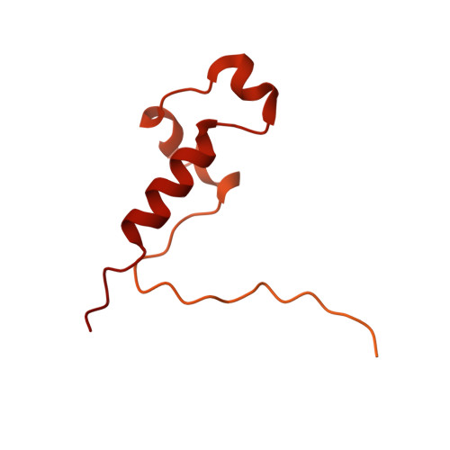
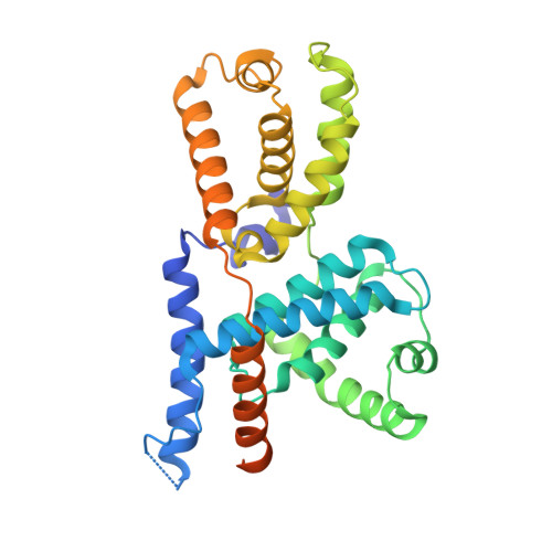
    The human CDK-activating kinase (CAK), a complex composed of cyclin-dependent kinase (CDK) 7, cyclin H, and MAT1, is a critical regulator of transcription initiation and the cell cycle. It acts by phosphorylating the C-terminal heptapeptide repeat domain of the RNA polymerase II (Pol II) subunit RPB1, which is an important regulatory event in transcription initiation by Pol II, and it phosphorylates the regulatory T-loop of CDKs that control cell cycle progression. Here, we have determined the three-dimensional (3D) structure of the catalytic module of human CAK, revealing the structural basis of its assembly and providing insight into CDK7 activation in this context. The unique third component of the complex, MAT1, substantially extends the interaction interface between CDK7 and cyclin H, explaining its role as a CAK assembly factor, and it forms interactions with the CDK7 T-loop, which may contribute to enhancing CAK activity. We have also determined the structure of the CAK in complex with the covalently bound inhibitor THZ1 in order to provide insight into the binding of inhibitors at the CDK7 active site and to aid in the rational design of therapeutic compounds.


  • Organizational Affiliation

    California Institute for Quantitative Biosciences (QB3), University of California, Berkeley, CA 94720; basil.greber@icr.ac.uk ENogales@lbl.gov.


Macromolecules
Find similar proteins by:  (by identity cutoff)  |  3D Structure
Entity ID: 1
MoleculeChains Sequence LengthOrganismDetailsImage
CDK-activating kinase assembly factor MAT1A [auth H]328Homo sapiensMutation(s): 0 
Gene Names: MNAT1CAP35MAT1RNF66
UniProt & NIH Common Fund Data Resources
Find proteins for P51948 (Homo sapiens)
Explore P51948 
Go to UniProtKB:  P51948
PHAROS:  P51948
GTEx:  ENSG00000020426 
Entity Groups  
Sequence Clusters30% Identity50% Identity70% Identity90% Identity95% Identity100% Identity
UniProt GroupP51948
Sequence Annotations
Expand
  • Reference Sequence
Find similar proteins by:  (by identity cutoff)  |  3D Structure
Entity ID: 2
MoleculeChains Sequence LengthOrganismDetailsImage
Cyclin-HB [auth I]323Homo sapiensMutation(s): 0 
Gene Names: CCNH
UniProt & NIH Common Fund Data Resources
Find proteins for P51946 (Homo sapiens)
Explore P51946 
Go to UniProtKB:  P51946
PHAROS:  P51946
GTEx:  ENSG00000134480 
Entity Groups  
Sequence Clusters30% Identity50% Identity70% Identity90% Identity95% Identity100% Identity
UniProt GroupP51946
Sequence Annotations
Expand
  • Reference Sequence
Find similar proteins by:  (by identity cutoff)  |  3D Structure
Entity ID: 3
MoleculeChains Sequence LengthOrganismDetailsImage
Cyclin-dependent kinase 7C [auth J]391Homo sapiensMutation(s): 0 
Gene Names: CDK7CAKCAK1CDKN7MO15STK1
EC: 2.7.11.22 (PDB Primary Data), 2.7.11.23 (PDB Primary Data)
UniProt & NIH Common Fund Data Resources
Find proteins for P50613 (Homo sapiens)
Explore P50613 
Go to UniProtKB:  P50613
PHAROS:  P50613
GTEx:  ENSG00000134058 
Entity Groups  
Sequence Clusters30% Identity50% Identity70% Identity90% Identity95% Identity100% Identity
UniProt GroupP50613
Sequence Annotations
Expand
  • Reference Sequence
Small Molecules
Ligands 2 Unique
IDChains Name / Formula / InChI Key2D Diagram3D Interactions
AGS
Query on AGS

Download Ideal Coordinates CCD File 
E [auth J]PHOSPHOTHIOPHOSPHORIC ACID-ADENYLATE ESTER
C10 H16 N5 O12 P3 S
NLTUCYMLOPLUHL-KQYNXXCUSA-N
MG
Query on MG

Download Ideal Coordinates CCD File 
D [auth J]MAGNESIUM ION
Mg
JLVVSXFLKOJNIY-UHFFFAOYSA-N
Modified Residues  1 Unique
IDChains TypeFormula2D DiagramParent
SEP
Query on SEP
C [auth J]L-PEPTIDE LINKINGC3 H8 N O6 PSER
Experimental Data & Validation

Experimental Data

  • Method: ELECTRON MICROSCOPY
  • Resolution: 2.80 Å
  • Aggregation State: PARTICLE 
  • Reconstruction Method: SINGLE PARTICLE 
EM Software:
TaskSoftware PackageVersion
RECONSTRUCTIONRELION3.1
MODEL REFINEMENTPHENIX

Structure Validation

View Full Validation Report


Entry History & Funding Information

Deposition Data


Funding OrganizationLocationGrant Number
National Institutes of Health/National Institute of General Medical Sciences (NIH/NIGMS)United StatesR01-GM63072
National Institutes of Health/National Institute of General Medical Sciences (NIH/NIGMS)United StatesP01-GM063210
National Institutes of Health/National Institute of General Medical Sciences (NIH/NIGMS)United StatesR35-GM127018
Howard Hughes Medical Institute (HHMI)United States--

Revision History  (Full details and data files)

  • Version 1.0: 2020-09-09
    Type: Initial release
  • Version 1.1: 2020-09-30
    Changes: Database references