6XAR

Structure of CBL tyrosine kinase binding domain (TKBD) with C-terminal tail of Src-like kinase protein 2 (SLAP2)


Experimental Data Snapshot

  • Method: X-RAY DIFFRACTION
  • Resolution: 2.50 Å
  • R-Value Free: 0.293 
  • R-Value Work: 0.249 
  • R-Value Observed: 0.252 

wwPDB Validation   3D Report Full Report


This is version 1.2 of the entry. See complete history


Literature

SLAP2 Adaptor Binding Disrupts c-CBL Autoinhibition to Activate Ubiquitin Ligase Function.

Wybenga-Groot, L.E.Tench, A.J.Simpson, C.D.Germain, J.S.Raught, B.Moran, M.F.McGlade, C.J.

(2021) J Mol Biol 433: 166880-166880

  • DOI: https://doi.org/10.1016/j.jmb.2021.166880
  • Primary Citation of Related Structures:  
    6XAR

  • PubMed Abstract: 

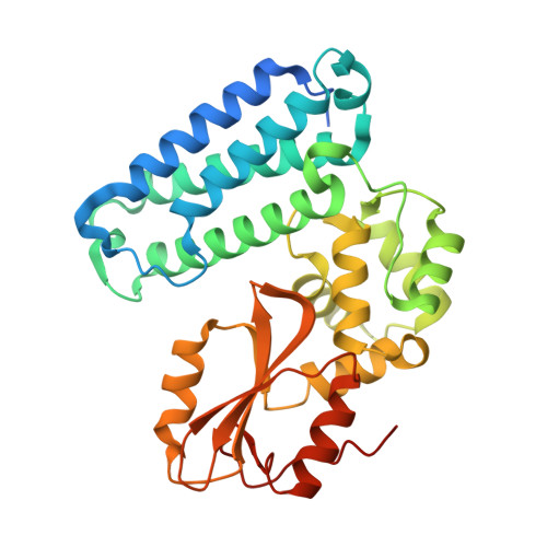
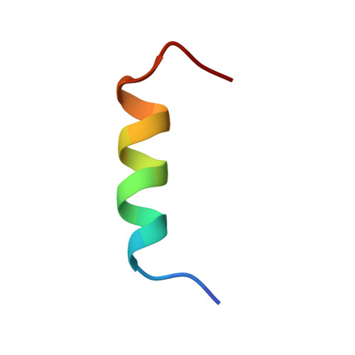
    CBL is a RING type E3 ubiquitin ligase that functions as a negative regulator of tyrosine kinase signaling and loss of CBL E3 function is implicated in several forms of leukemia. The Src-like adaptor proteins (SLAP/SLAP2) bind to CBL and are required for CBL-dependent downregulation of antigen receptor, cytokine receptor, and receptor tyrosine kinase signaling. Despite the established role of SLAP/SLAP2 in regulating CBL activity, the nature of the interaction and the mechanisms involved are not known. To understand the molecular basis of the interaction between SLAP/SLAP2 and CBL, we solved the crystal structure of CBL tyrosine kinase binding domain (TKBD) in complex with SLAP2. The carboxy-terminal region of SLAP2 adopts an α-helical structure which binds in a cleft between the 4H, EF-hand, and SH2 domains of the TKBD. This SLAP2 binding site is remote from the canonical TKBD phospho-tyrosine peptide binding site but overlaps with a region important for stabilizing CBL in its autoinhibited conformation. In addition, binding of SLAP2 to CBL in vitro activates the ubiquitin ligase function of autoinhibited CBL. Disruption of the CBL/SLAP2 interface through mutagenesis demonstrated a role for this protein-protein interaction in regulation of CBL E3 ligase activity in cells. Our results reveal that SLAP2 binding to a regulatory cleft of the TKBD provides an alternative mechanism for activation of CBL ubiquitin ligase function.


  • Organizational Affiliation

    The Arthur and Sonia Labatt Brain Tumour Research Centre, The Hospital for Sick Children, 555 University Avenue, Toronto, ON M5G 1X8, Canada; Program in Cell Biology, The Hospital for Sick Children, 555 University Avenue, Toronto, ON M5G 1X8, Canada; SPARC BioCentre, The Hospital for Sick Children, 555 University Avenue, Toronto, ON M5G 1X8, Canada. Electronic address: leanne.wybenga-groot@sickkids.ca.


Macromolecules
Find similar proteins by:  (by identity cutoff)  |  3D Structure
Entity ID: 1
MoleculeChains Sequence LengthOrganismDetailsImage
E3 ubiquitin-protein ligase CBL
A, B
335Homo sapiensMutation(s): 0 
Gene Names: CBLCBL2RNF55
EC: 2.3.2.27
UniProt & NIH Common Fund Data Resources
Find proteins for P22681 (Homo sapiens)
Explore P22681 
Go to UniProtKB:  P22681
PHAROS:  P22681
GTEx:  ENSG00000110395 
Entity Groups  
Sequence Clusters30% Identity50% Identity70% Identity90% Identity95% Identity100% Identity
UniProt GroupP22681
Sequence Annotations
Expand
  • Reference Sequence

Find similar proteins by:  Sequence   |   3D Structure  

Entity ID: 2
MoleculeChains Sequence LengthOrganismDetailsImage
Src-like-adapter 2
C, D
19Mus musculusMutation(s): 0 
Gene Names: Sla2
UniProt & NIH Common Fund Data Resources
Find proteins for Q8R4L0 (Mus musculus)
Explore Q8R4L0 
Go to UniProtKB:  Q8R4L0
IMPC:  MGI:1925049
Entity Groups  
Sequence Clusters30% Identity50% Identity70% Identity90% Identity95% Identity100% Identity
UniProt GroupQ8R4L0
Sequence Annotations
Expand
  • Reference Sequence
Experimental Data & Validation

Experimental Data

  • Method: X-RAY DIFFRACTION
  • Resolution: 2.50 Å
  • R-Value Free: 0.293 
  • R-Value Work: 0.249 
  • R-Value Observed: 0.252 
  • Space Group: P 1 21 1
Unit Cell:
Length ( Å )Angle ( ˚ )
a = 62.73α = 90
b = 86.96β = 112.31
c = 65.26γ = 90
Software Package:
Software NamePurpose
MOSFLMdata reduction
Aimlessdata scaling
PHASERphasing
PHENIXrefinement
PDB_EXTRACTdata extraction

Structure Validation

View Full Validation Report


Entry History & Funding Information

Deposition Data


Funding OrganizationLocationGrant Number
Canadian Institutes of Health Research (CIHR)CanadaMOP-12859
Canadian Institutes of Health Research (CIHR)CanadaFRN166034

Revision History  (Full details and data files)

  • Version 1.0: 2021-03-03
    Type: Initial release
  • Version 1.1: 2021-10-06
    Changes: Database references
  • Version 1.2: 2023-10-18
    Changes: Data collection, Refinement description