6WIC

Pre-catalytic quaternary complex of human Polymerase Mu on a complementary DNA double-strand break substrate


Experimental Data Snapshot

  • Method: X-RAY DIFFRACTION
  • Resolution: 1.55 Å
  • R-Value Free: 0.179 
  • R-Value Work: 0.166 
  • R-Value Observed: 0.166 

wwPDB Validation   3D Report Full Report


Ligand Structure Quality Assessment 


This is version 1.1 of the entry. See complete history


Literature

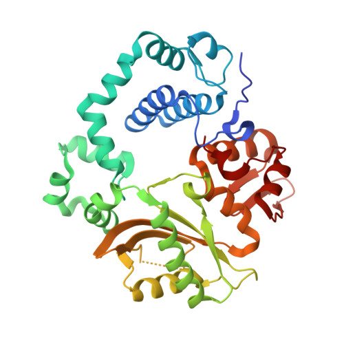
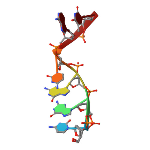
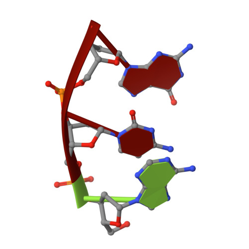
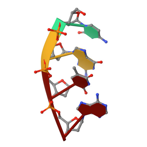
Structural snapshots of human DNA polymerase mu engaged on a DNA double-strand break.

Kaminski, A.M.Pryor, J.M.Ramsden, D.A.Kunkel, T.A.Pedersen, L.C.Bebenek, K.

(2020) Nat Commun 11: 4784-4784

  • DOI: https://doi.org/10.1038/s41467-020-18506-5
  • Primary Citation of Related Structures:  
    6WIC, 6WID, 6WIE

  • PubMed Abstract: 

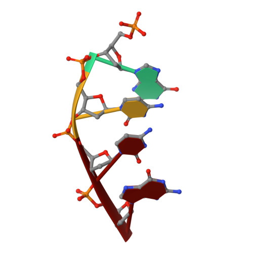
    Genomic integrity is threatened by cytotoxic DNA double-strand breaks (DSBs), which must be resolved efficiently to prevent sequence loss, chromosomal rearrangements/translocations, or cell death. Polymerase μ (Polμ) participates in DSB repair via the nonhomologous end-joining (NHEJ) pathway, by filling small sequence gaps in broken ends to create substrates ultimately ligatable by DNA Ligase IV. Here we present structures of human Polμ engaging a DSB substrate. Synapsis is mediated solely by Polμ, facilitated by single-nucleotide homology at the break site, wherein both ends of the discontinuous template strand are stabilized by a hydrogen bonding network. The active site in the quaternary Pol μ complex is poised for catalysis and nucleotide incoporation proceeds in crystallo. These structures demonstrate that Polμ may address complementary DSB substrates during NHEJ in a manner indistinguishable from single-strand breaks.


  • Organizational Affiliation

    Genome Integrity and Structural Biology Laboratory, National Institute of Environmental Health Sciences, National Institutes of Health, 111 TW Alexander Dr., Bldg. 101/Rm F338, Research Triangle Park, NC, 27709, USA.


Macromolecules

Find similar proteins by:  (by identity cutoff)  |  3D Structure
Entity ID: 1
MoleculeChains Sequence LengthOrganismDetailsImage
DNA-directed DNA/RNA polymerase mu356Homo sapiensMutation(s): 0 
Gene Names: POLMpolmu
EC: 2.7.7.7
UniProt & NIH Common Fund Data Resources
Find proteins for Q9NP87 (Homo sapiens)
Explore Q9NP87 
Go to UniProtKB:  Q9NP87
PHAROS:  Q9NP87
GTEx:  ENSG00000122678 
Entity Groups  
Sequence Clusters30% Identity50% Identity70% Identity90% Identity95% Identity100% Identity
UniProt GroupQ9NP87
Sequence Annotations
Expand
  • Reference Sequence

Find similar nucleic acids by:  Sequence   |   3D Structure  

Entity ID: 2
MoleculeChains LengthOrganismImage
DNA (5'-D(*CP*GP*GP*CP*AP*T)-3')B [auth T]6synthetic construct
Sequence Annotations
Expand
  • Reference Sequence

Find similar nucleic acids by:  Sequence   |   3D Structure  

Entity ID: 3
MoleculeChains LengthOrganismImage
DNA (5'-D(*AP*CP*G)-3')C [auth U]3synthetic construct
Sequence Annotations
Expand
  • Reference Sequence

Find similar nucleic acids by:  Sequence   |   3D Structure  

Entity ID: 4
MoleculeChains LengthOrganismImage
DNA (5'-D(*CP*GP*TP*A)-3')D [auth P]4synthetic construct
Sequence Annotations
Expand
  • Reference Sequence

Find similar nucleic acids by:  Sequence   |   3D Structure  

Entity ID: 5
MoleculeChains LengthOrganismImage
DNA (5'-D(P*GP*CP*CP*G)-3')E [auth D]4synthetic construct
Sequence Annotations
Expand
  • Reference Sequence
Small Molecules
Ligands 8 Unique
IDChains Name / Formula / InChI Key2D Diagram3D Interactions
DUP (Subject of Investigation/LOI)
Query on DUP

Download Ideal Coordinates CCD File 
F [auth A]2'-DEOXYURIDINE 5'-ALPHA,BETA-IMIDO-TRIPHOSPHATE
C9 H16 N3 O13 P3
XZLLMTSKYYYJLH-SHYZEUOFSA-N
1PE
Query on 1PE

Download Ideal Coordinates CCD File 
N [auth A]PENTAETHYLENE GLYCOL
C10 H22 O6
JLFNLZLINWHATN-UHFFFAOYSA-N
PEG
Query on PEG

Download Ideal Coordinates CCD File 
M [auth A]DI(HYDROXYETHYL)ETHER
C4 H10 O3
MTHSVFCYNBDYFN-UHFFFAOYSA-N
GOL
Query on GOL

Download Ideal Coordinates CCD File 
L [auth A]GLYCEROL
C3 H8 O3
PEDCQBHIVMGVHV-UHFFFAOYSA-N
K
Query on K

Download Ideal Coordinates CCD File 
J [auth A]POTASSIUM ION
K
NPYPAHLBTDXSSS-UHFFFAOYSA-N
CL
Query on CL

Download Ideal Coordinates CCD File 
K [auth A]CHLORIDE ION
Cl
VEXZGXHMUGYJMC-UHFFFAOYSA-M
MG (Subject of Investigation/LOI)
Query on MG

Download Ideal Coordinates CCD File 
G [auth A],
H [auth A]
MAGNESIUM ION
Mg
JLVVSXFLKOJNIY-UHFFFAOYSA-N
NA
Query on NA

Download Ideal Coordinates CCD File 
I [auth A]SODIUM ION
Na
FKNQFGJONOIPTF-UHFFFAOYSA-N
Experimental Data & Validation

Experimental Data

  • Method: X-RAY DIFFRACTION
  • Resolution: 1.55 Å
  • R-Value Free: 0.179 
  • R-Value Work: 0.166 
  • R-Value Observed: 0.166 
  • Space Group: P 21 21 21
Unit Cell:
Length ( Å )Angle ( ˚ )
a = 60.134α = 90
b = 62.233β = 90
c = 118.054γ = 90
Software Package:
Software NamePurpose
PHENIXrefinement
HKL-2000data reduction
HKL-2000data scaling
PHASERphasing

Structure Validation

View Full Validation Report


Ligand Structure Quality Assessment 


Entry History & Funding Information

Deposition Data


Funding OrganizationLocationGrant Number
National Institutes of Health/National Institute of Environmental Health Sciences (NIH/NIEHS)United States1ZIA ES 102645
National Institutes of Health/National Institute of Environmental Health Sciences (NIH/NIEHS)United StatesZ01 ES065070

Revision History  (Full details and data files)

  • Version 1.0: 2020-10-07
    Type: Initial release
  • Version 1.1: 2023-10-18
    Changes: Data collection, Database references, Refinement description