6V0Z

Structure of ALDH7A1 mutant R441C complexed with NAD


Experimental Data Snapshot

  • Method: X-RAY DIFFRACTION
  • Resolution: 2.02 Å
  • R-Value Free: 0.249 
  • R-Value Work: 0.194 
  • R-Value Observed: 0.196 

wwPDB Validation   3D Report Full Report

Currently 6V0Z does not have a validation slider image.


This is version 1.2 of the entry. See complete history


Literature

Biochemical, structural, and computational analyses of two new clinically identified missense mutations of ALDH7A1.

Korasick, D.A.Buckley, D.P.Palpacelli, A.Cursio, I.Cesaroni, E.Cheng, J.Tanner, J.J.

(2024) Chem Biol Interact : 110993-110993

  • DOI: https://doi.org/10.1016/j.cbi.2024.110993
  • Primary Citation of Related Structures:  
    6V0Z

  • PubMed Abstract: 

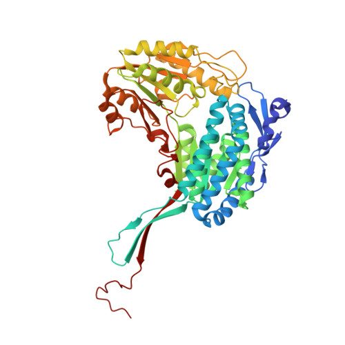
    Aldehyde dehydrogenase 7A1 (ALDH7A1) catalyzes a step of lysine catabolism. Certain missense mutations in the ALDH7A1 gene cause pyridoxine dependent epilepsy (PDE), a rare autosomal neurometabolic disorder with recessive inheritance that affects almost 1:65,000 live births and is classically characterized by recurrent seizures from the neonatal period. We report a biochemical, structural, and computational study of two novel ALDH7A1 missense mutations that were identified in a child with rare recurrent seizures from the third month of life. The mutations affect two residues in the oligomer interfaces of ALDH7A1, Arg134 and Arg441 (Arg162 and Arg469 in the HGVS nomenclature). The corresponding enzyme variants R134S and R441C (p.Arg162Ser and p.Arg469Cys in the HGVS nomenclature) were expressed in Escherichia coli and purified. R134S and R441C have 10,000- and 50-fold lower catalytic efficiency than wild-type ALDH7A1, respectively. Sedimentation velocity analytical ultracentrifugation shows that R134S is defective in tetramerization, remaining locked in a dimeric state even in the presence of the tetramer-inducing coenzyme NAD + . Because the tetramer is the active form of ALDH7A1, the defect in oligomerization explains the very low catalytic activity of R134S. In contrast, R441C exhibits wild-type oligomerization behavior, and the 2.0 Å resolution crystal structure of R441C complexed with NAD + revealed no obvious structural perturbations when compared to the wild-type enzyme structure. Molecular dynamics simulations suggest that the mutation of Arg441 to Cys may increase intersubunit ion pairs and alter the dynamics of the active site gate. Our biochemical, structural, and computational data on two novel clinical variants of ALDH7A1 add to the complexity of the molecular determinants underlying pyridoxine dependent epilepsy.


  • Organizational Affiliation

    Department of Biochemistry, University of Missouri, Columbia, MO, 65211, United States.


Macromolecules
Find similar proteins by:  (by identity cutoff)  |  3D Structure
Entity ID: 1
MoleculeChains Sequence LengthOrganismDetailsImage
Alpha-aminoadipic semialdehyde dehydrogenase
A, B, C, D, E
A, B, C, D, E, F, G, H
513Homo sapiensMutation(s): 1 
Gene Names: ALDH7A1ATQ1
EC: 1.2.1.31 (PDB Primary Data), 1.2.1.3 (PDB Primary Data), 1.2.1.8 (PDB Primary Data)
UniProt & NIH Common Fund Data Resources
Find proteins for P49419 (Homo sapiens)
Explore P49419 
Go to UniProtKB:  P49419
PHAROS:  P49419
GTEx:  ENSG00000164904 
Entity Groups  
Sequence Clusters30% Identity50% Identity70% Identity90% Identity95% Identity100% Identity
UniProt GroupP49419
Sequence Annotations
Expand
  • Reference Sequence
Small Molecules
Ligands 2 Unique
IDChains Name / Formula / InChI Key2D Diagram3D Interactions
NAD (Subject of Investigation/LOI)
Query on NAD

Download Ideal Coordinates CCD File 
I [auth A]
J [auth B]
K [auth C]
M [auth D]
N [auth E]
I [auth A],
J [auth B],
K [auth C],
M [auth D],
N [auth E],
O [auth F],
P [auth G],
Q [auth H]
NICOTINAMIDE-ADENINE-DINUCLEOTIDE
C21 H27 N7 O14 P2
BAWFJGJZGIEFAR-NNYOXOHSSA-N
EDO
Query on EDO

Download Ideal Coordinates CCD File 
L [auth C]1,2-ETHANEDIOL
C2 H6 O2
LYCAIKOWRPUZTN-UHFFFAOYSA-N
Experimental Data & Validation

Experimental Data

  • Method: X-RAY DIFFRACTION
  • Resolution: 2.02 Å
  • R-Value Free: 0.249 
  • R-Value Work: 0.194 
  • R-Value Observed: 0.196 
  • Space Group: C 1 2 1
Unit Cell:
Length ( Å )Angle ( ˚ )
a = 154.964α = 90
b = 161.647β = 94.45
c = 158.542γ = 90
Software Package:
Software NamePurpose
Aimlessdata scaling
PHENIXrefinement
PDB_EXTRACTdata extraction
XDSdata reduction
PHASERphasing

Structure Validation

View Full Validation Report

Currently 6V0Z does not have a validation slider image.


Entry History & Funding Information

Deposition Data


Funding OrganizationLocationGrant Number
National Institutes of Health/National Institute of General Medical Sciences (NIH/NIGMS)United StatesR01GM093123

Revision History  (Full details and data files)

  • Version 1.0: 2020-11-25
    Type: Initial release
  • Version 1.1: 2023-10-11
    Changes: Data collection, Database references, Refinement description
  • Version 1.2: 2024-04-24
    Changes: Database references