6TCA

Phosphorylated p38 and MAPKAPK2 complex with inhibitor


Experimental Data Snapshot

  • Method: X-RAY DIFFRACTION
  • Resolution: 3.70 Å
  • R-Value Free: 0.297 
  • R-Value Work: 0.251 
  • R-Value Observed: 0.254 

wwPDB Validation   3D Report Full Report


Ligand Structure Quality Assessment 


This is version 1.2 of the entry. See complete history


Literature

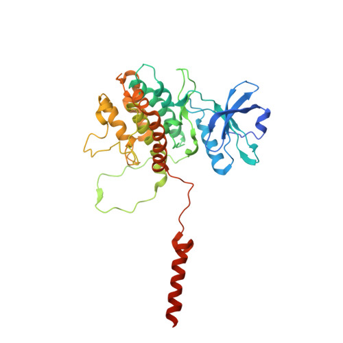
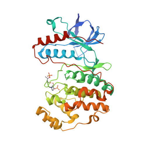
MAP Kinase-Mediated Activation of RSK1 and MK2 Substrate Kinases.

Sok, P.Gogl, G.Kumar, G.S.Alexa, A.Singh, N.Kirsch, K.Sebo, A.Drahos, L.Gaspari, Z.Peti, W.Remenyi, A.

(2020) Structure 28: 1101-1113.e5

  • DOI: https://doi.org/10.1016/j.str.2020.06.007
  • Primary Citation of Related Structures:  
    6TCA

  • PubMed Abstract: 

    Mitogen-activated protein kinases (MAPKs) control essential eukaryotic signaling pathways. While much has been learned about MAPK activation, much less is known about substrate recruitment and specificity. MAPK substrates may be other kinases that are crucial to promote a further diversification of the signaling outcomes. Here, we used a variety of molecular and cellular tools to investigate the recruitment of two substrate kinases, RSK1 and MK2, to three MAPKs (ERK2, p38α, and ERK5). Unexpectedly, we identified that kinase heterodimers form structurally and functionally distinct complexes depending on the activation state of the MAPK. These may be incompatible with downstream signaling, but naturally they may also form structures that are compatible with the phosphorylation of the downstream kinase at the activation loop, or alternatively at other allosteric sites. Furthermore, we show that small-molecule inhibitors may affect the quaternary arrangement of kinase heterodimers and thus influence downstream signaling in a specific manner.


  • Organizational Affiliation

    Biomolecular Interactions Research Group, Institute of Organic Chemistry, Research Center for Natural Sciences, Magyar Tudósok körútja 2., 1117 Budapest, Hungary.


Macromolecules
Find similar proteins by:  (by identity cutoff)  |  3D Structure
Entity ID: 1
MoleculeChains Sequence LengthOrganismDetailsImage
MAP kinase-activated protein kinase 2
A, C, E, G
371Homo sapiensMutation(s): 0 
Gene Names: MAPKAPK2
EC: 2.7.11.1
UniProt & NIH Common Fund Data Resources
Find proteins for P49137 (Homo sapiens)
Explore P49137 
Go to UniProtKB:  P49137
PHAROS:  P49137
GTEx:  ENSG00000162889 
Entity Groups  
Sequence Clusters30% Identity50% Identity70% Identity90% Identity95% Identity100% Identity
UniProt GroupP49137
Sequence Annotations
Expand
  • Reference Sequence
Find similar proteins by:  (by identity cutoff)  |  3D Structure
Entity ID: 2
MoleculeChains Sequence LengthOrganismDetailsImage
Mitogen-activated protein kinase 14
B, D, F, H
364Homo sapiensMutation(s): 0 
Gene Names: MAPK14CSBPCSBP1CSBP2CSPB1MXI2SAPK2A
EC: 2.7.11.24
UniProt & NIH Common Fund Data Resources
Find proteins for Q16539 (Homo sapiens)
Explore Q16539 
Go to UniProtKB:  Q16539
PHAROS:  Q16539
GTEx:  ENSG00000112062 
Entity Groups  
Sequence Clusters30% Identity50% Identity70% Identity90% Identity95% Identity100% Identity
UniProt GroupQ16539
Sequence Annotations
Expand
  • Reference Sequence
Small Molecules
Modified Residues  2 Unique
IDChains TypeFormula2D DiagramParent
PTR
Query on PTR
B, D, F, H
L-PEPTIDE LINKINGC9 H12 N O6 PTYR
TPO
Query on TPO
B, D, F, H
L-PEPTIDE LINKINGC4 H10 N O6 PTHR
Experimental Data & Validation

Experimental Data

  • Method: X-RAY DIFFRACTION
  • Resolution: 3.70 Å
  • R-Value Free: 0.297 
  • R-Value Work: 0.251 
  • R-Value Observed: 0.254 
  • Space Group: C 1 2 1
Unit Cell:
Length ( Å )Angle ( ˚ )
a = 279.346α = 90
b = 69.286β = 123.85
c = 221.107γ = 90
Software Package:
Software NamePurpose
XDSdata reduction
Aimlessdata scaling
PHASERphasing
PHENIXrefinement
PDB_EXTRACTdata extraction

Structure Validation

View Full Validation Report


Ligand Structure Quality Assessment 


Entry History & Funding Information

Deposition Data


Funding OrganizationLocationGrant Number
European UnionHungaryiNEXT: PID 1742, PID 4788

Revision History  (Full details and data files)

  • Version 1.0: 2020-07-22
    Type: Initial release
  • Version 1.1: 2020-10-14
    Changes: Database references
  • Version 1.2: 2024-01-24
    Changes: Data collection, Database references, Refinement description