6T9O

CryoEM structure of human polycystin-2/PKD2 in UDM supplemented with PI(3,5)P2


Experimental Data Snapshot

  • Method: ELECTRON MICROSCOPY
  • Resolution: 3.39 Å
  • Aggregation State: PARTICLE 
  • Reconstruction Method: SINGLE PARTICLE 

wwPDB Validation   3D Report Full Report


This is version 2.0 of the entry. See complete history


Literature

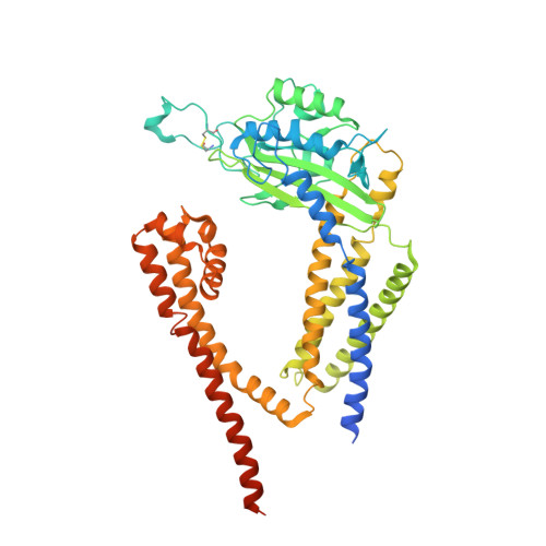
Lipid Interactions of a Ciliary Membrane TRP Channel: Simulation and Structural Studies of Polycystin-2.

Wang, Q.Corey, R.A.Hedger, G.Aryal, P.Grieben, M.Nasrallah, C.Baronina, A.Pike, A.C.W.Shi, J.Carpenter, E.P.Sansom, M.S.P.

(2020) Structure 28: 169-184.e5

  • DOI: https://doi.org/10.1016/j.str.2019.11.005
  • Primary Citation of Related Structures:  
    6T9N, 6T9O

  • PubMed Abstract: 

    Polycystin-2 (PC2) is a transient receptor potential (TRP) channel present in ciliary membranes of the kidney. PC2 shares a transmembrane fold with other TRP channels, in addition to an extracellular domain found in TRPP and TRPML channels. Using molecular dynamics (MD) simulations and cryoelectron microscopy we identify and characterize PIP 2 and cholesterol interactions with PC2. PC2 is revealed to have a PIP binding site close to the equivalent vanilloid/lipid binding site in the TRPV1 channel. A 3.0-Å structure reveals a binding site for cholesterol on PC2. Cholesterol interactions with the channel at this site are characterized by MD simulations. The two classes of lipid binding sites are compared with sites observed in other TRPs and in Kv channels. These findings suggest PC2, in common with other ion channels, may be modulated by both PIPs and cholesterol, and position PC2 within an emerging model of the roles of lipids in the regulation and organization of ciliary membranes.


  • Organizational Affiliation

    Department of Biochemistry, University of Oxford, South Parks Road, Oxford OX1 3QU, UK; Structural Genomics Consortium, University of Oxford, Old Road Campus Research Building, Roosevelt Drive, Oxford OX3 7DQ, UK.


Macromolecules
Find similar proteins by:  (by identity cutoff)  |  3D Structure
Entity ID: 1
MoleculeChains Sequence LengthOrganismDetailsImage
Polycystin-2
A, B, C, D
547Homo sapiensMutation(s): 0 
Gene Names: PKD2TRPP2
Membrane Entity: Yes 
UniProt & NIH Common Fund Data Resources
Find proteins for Q13563 (Homo sapiens)
Explore Q13563 
Go to UniProtKB:  Q13563
PHAROS:  Q13563
GTEx:  ENSG00000118762 
Entity Groups  
Sequence Clusters30% Identity50% Identity70% Identity90% Identity95% Identity100% Identity
UniProt GroupQ13563
Sequence Annotations
Expand
  • Reference Sequence
Oligosaccharides

Help

Entity ID: 2
MoleculeChains Length2D Diagram Glycosylation3D Interactions
2-acetamido-2-deoxy-beta-D-glucopyranose-(1-4)-2-acetamido-2-deoxy-beta-D-glucopyranose
E, F, G, H
2N-Glycosylation
Glycosylation Resources
GlyTouCan:  G42666HT
GlyCosmos:  G42666HT
GlyGen:  G42666HT
Small Molecules
Ligands 4 Unique
IDChains Name / Formula / InChI Key2D Diagram3D Interactions
UMQ
Query on UMQ

Download Ideal Coordinates CCD File 
CA [auth C]
DA [auth C]
EA [auth C]
FA [auth C]
GA [auth C]
CA [auth C],
DA [auth C],
EA [auth C],
FA [auth C],
GA [auth C],
KA [auth D],
L [auth A],
LA [auth D],
M [auth A],
MA [auth D],
N [auth A],
NA [auth D],
O [auth A],
OA [auth D],
P [auth A],
U [auth B],
V [auth B],
W [auth B],
X [auth B],
Y [auth B]
UNDECYL-MALTOSIDE
C23 H44 O11
UYEMNFYVTFDKRG-ZNGNCRBCSA-N
CLR
Query on CLR

Download Ideal Coordinates CCD File 
BA [auth C],
JA [auth D],
K [auth A],
T [auth B]
CHOLESTEROL
C27 H46 O
HVYWMOMLDIMFJA-DPAQBDIFSA-N
NAG
Query on NAG

Download Ideal Coordinates CCD File 
AA [auth C]
HA [auth D]
I [auth A]
IA [auth D]
J [auth A]
AA [auth C],
HA [auth D],
I [auth A],
IA [auth D],
J [auth A],
R [auth B],
S [auth B],
Z [auth C]
2-acetamido-2-deoxy-beta-D-glucopyranose
C8 H15 N O6
OVRNDRQMDRJTHS-FMDGEEDCSA-N
CA
Query on CA

Download Ideal Coordinates CCD File 
Q [auth A]CALCIUM ION
Ca
BHPQYMZQTOCNFJ-UHFFFAOYSA-N
Experimental Data & Validation

Experimental Data

  • Method: ELECTRON MICROSCOPY
  • Resolution: 3.39 Å
  • Aggregation State: PARTICLE 
  • Reconstruction Method: SINGLE PARTICLE 
EM Software:
TaskSoftware PackageVersion
RECONSTRUCTIONRELION3
MODEL REFINEMENTPHENIX1.13

Structure Validation

View Full Validation Report


Entry History & Funding Information

Deposition Data


Funding OrganizationLocationGrant Number
Wellcome TrustUnited Kingdom106169/Z/14/Z

Revision History  (Full details and data files)

  • Version 1.0: 2019-11-20
    Type: Initial release
  • Version 1.1: 2019-12-18
    Changes: Database references
  • Version 1.2: 2020-02-12
    Changes: Data collection, Database references
  • Version 2.0: 2020-07-29
    Type: Remediation
    Reason: Carbohydrate remediation
    Changes: Advisory, Atomic model, Data collection, Derived calculations, Structure summary