6SAD

Structure of 14-3-3 gamma in complex with double phosphorylated caspase-2 peptide on Ser139 and Ser164


Experimental Data Snapshot

  • Method: X-RAY DIFFRACTION
  • Resolution: 2.75 Å
  • R-Value Free: 0.271 
  • R-Value Work: 0.230 
  • R-Value Observed: 0.232 

wwPDB Validation   3D Report Full Report


This is version 1.3 of the entry. See complete history


Literature

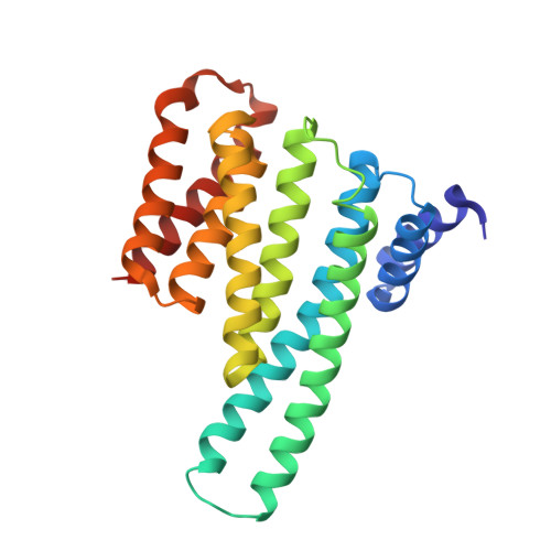
14-3-3 protein binding blocks the dimerization interface of caspase-2.

Kalabova, D.Filandr, F.Alblova, M.Petrvalska, O.Horvath, M.Man, P.Obsil, T.Obsilova, V.

(2020) FEBS J 287: 3494-3510

  • DOI: https://doi.org/10.1111/febs.15215
  • Primary Citation of Related Structures:  
    6S9K, 6SAD

  • PubMed Abstract: 

    Among all species, caspase-2 (C2) is the most evolutionarily conserved caspase required for effective initiation of apoptosis following death stimuli. C2 is activated through dimerization and autoproteolytic cleavage and inhibited through phosphorylation at Ser 139 and Ser 164 , within the linker between the caspase recruitment and p19 domains of the zymogen, followed by association with the adaptor protein 14-3-3, which maintains C2 in its immature form procaspase (proC2). However, the mechanism of 14-3-3-dependent inhibition of C2 activation remains unclear. Here, we report the structural characterization of the complex between proC2 and 14-3-3 by hydrogen/deuterium mass spectrometry and protein crystallography to determine the molecular basis for 14-3-3-mediated inhibition of C2 activation. Our data reveal that the 14-3-3 dimer interacts with proC2 not only through ligand-binding grooves but also through other regions outside the central channel, thus explaining the isoform-dependent specificity of 14-3-3 protein binding to proC2 and the substantially higher binding affinity of 14-3-3 protein to proC2 than to the doubly phosphorylated peptide. The formation of the complex between 14-3-3 protein and proC2 does not induce any large conformational change in proC2. Furthermore, 14-3-3 protein interacts with and masks both the nuclear localization sequence and the C-terminal region of the p12 domain of proC2 through transient interactions in which both the p19 and p12 domains of proC2 are not firmly docked onto the surface of 14-3-3. This masked region of p12 domain is involved in C2 dimerization. Therefore, 14-3-3 protein likely inhibits proC2 activation by blocking its dimerization surface. DATABASES: Structural data are available in the Protein Data Bank under the accession numbers 6SAD and 6S9K.


  • Organizational Affiliation

    Division BIOCEV, Department of Structural Biology of Signaling Proteins, Institute of Physiology of the Czech Academy of Sciences, Vestec, Czech Republic.


Macromolecules
Find similar proteins by:  (by identity cutoff)  |  3D Structure
Entity ID: 1
MoleculeChains Sequence LengthOrganismDetailsImage
14-3-3 protein gamma
A, B
234Homo sapiensMutation(s): 0 
Gene Names: YWHAG
UniProt & NIH Common Fund Data Resources
Find proteins for P61981 (Homo sapiens)
Explore P61981 
Go to UniProtKB:  P61981
PHAROS:  P61981
GTEx:  ENSG00000170027 
Entity Groups  
Sequence Clusters30% Identity50% Identity70% Identity90% Identity95% Identity100% Identity
UniProt GroupP61981
Sequence Annotations
Expand
  • Reference Sequence
Find similar proteins by:  (by identity cutoff)  |  3D Structure
Entity ID: 2
MoleculeChains Sequence LengthOrganismDetailsImage
Caspase-234Homo sapiensMutation(s): 0 
EC: 3.4.22.55
UniProt & NIH Common Fund Data Resources
Find proteins for P42575 (Homo sapiens)
Explore P42575 
Go to UniProtKB:  P42575
PHAROS:  P42575
GTEx:  ENSG00000106144 
Entity Groups  
Sequence Clusters30% Identity50% Identity70% Identity90% Identity95% Identity100% Identity
UniProt GroupP42575
Sequence Annotations
Expand
  • Reference Sequence
Small Molecules
Modified Residues  1 Unique
IDChains TypeFormula2D DiagramParent
SEP
Query on SEP
C
L-PEPTIDE LINKINGC3 H8 N O6 PSER
Experimental Data & Validation

Experimental Data

  • Method: X-RAY DIFFRACTION
  • Resolution: 2.75 Å
  • R-Value Free: 0.271 
  • R-Value Work: 0.230 
  • R-Value Observed: 0.232 
  • Space Group: P 63 2 2
Unit Cell:
Length ( Å )Angle ( ˚ )
a = 227.425α = 90
b = 227.425β = 90
c = 73.335γ = 120
Software Package:
Software NamePurpose
PHENIXrefinement
XDSdata reduction
XDSdata scaling
MOLREPphasing

Structure Validation

View Full Validation Report


Entry History & Funding Information

Deposition Data


Funding OrganizationLocationGrant Number
Czech Science FoundationCzech Republic17-00726S

Revision History  (Full details and data files)

  • Version 1.0: 2020-01-22
    Type: Initial release
  • Version 1.1: 2020-02-05
    Changes: Database references
  • Version 1.2: 2020-08-26
    Changes: Database references
  • Version 1.3: 2024-01-24
    Changes: Data collection, Database references, Refinement description