6QVL

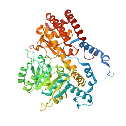
Human SHMT2 in complex with pemetrexed


Experimental Data Snapshot

  • Method: X-RAY DIFFRACTION
  • Resolution: 2.28 Å
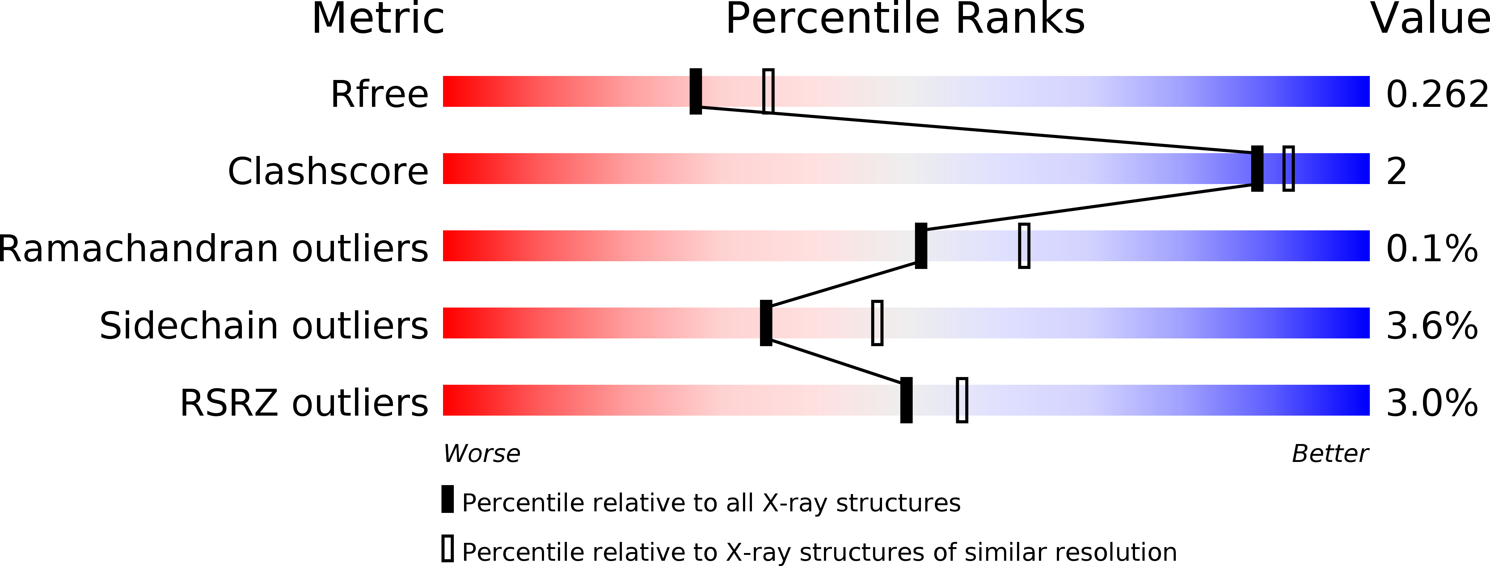
  • R-Value Free: 0.259 
  • R-Value Work: 0.212 
  • R-Value Observed: 0.214 

wwPDB Validation   3D Report Full Report


Ligand Structure Quality Assessment 


This is version 1.0 of the entry. See complete history


Literature

Structural basis of inhibition of the human serine hydroxymethyltransferase SHMT2 by antifolate drugs.

Scaletti, E.Jemth, A.S.Helleday, T.Stenmark, P.

(2019) FEBS Lett 593: 1863-1873

  • DOI: https://doi.org/10.1002/1873-3468.13455
  • Primary Citation of Related Structures:  
    6QVG, 6QVL

  • PubMed Abstract: 

    Serine hydroxymethyltransferase (SHMT) is the major source of 1-carbon units required for nucleotide synthesis. Humans have cytosolic (SHMT1) and mitochondrial (SHMT2) isoforms, which are upregulated in numerous cancers, making the enzyme an attractive drug target. Here, we show that the antifolates lometrexol and pemetrexed are inhibitors of SHMT2 and solve the first SHMT2-antifolate structures. The antifolates display large differences in their hydrogen bond networks despite their similarity. Lometrexol was found to be the best hSHMT1/2 inhibitor from a panel antifolates. Comparison of apo hSHMT1 with antifolate bound hSHMT2 indicates a highly conserved active site architecture. This structural information offers insights as to how these compounds could be improved to produce more potent and specific inhibitors of this emerging anti-cancer drug target.


  • Organizational Affiliation

    Department of Biochemistry and Biophysics, Stockholm University, Sweden.


Macromolecules
Find similar proteins by:  (by identity cutoff)  |  3D Structure
Entity ID: 1
MoleculeChains Sequence LengthOrganismDetailsImage
Serine hydroxymethyltransferase, mitochondrial
A, B
504Homo sapiensMutation(s): 0 
Gene Names: SHMT2
EC: 2.1.2.1
UniProt & NIH Common Fund Data Resources
Find proteins for P34897 (Homo sapiens)
Explore P34897 
Go to UniProtKB:  P34897
PHAROS:  P34897
GTEx:  ENSG00000182199 
Entity Groups  
Sequence Clusters30% Identity50% Identity70% Identity90% Identity95% Identity100% Identity
UniProt GroupP34897
Sequence Annotations
Expand
  • Reference Sequence
Small Molecules
Ligands 5 Unique
IDChains Name / Formula / InChI Key2D Diagram3D Interactions
4DW (Subject of Investigation/LOI)
Query on 4DW

Download Ideal Coordinates CCD File 
I [auth B]N-{4-[2-(2-amino-4-oxo-4,7-dihydro-3H-pyrrolo[2,3-d]pyrimidin-5-yl)ethyl]benzoyl}-L-glutamic acid
C20 H19 N5 O6
OOPGILDDKAKTLT-GWJCSSMESA-N
PLP
Query on PLP

Download Ideal Coordinates CCD File 
G [auth A],
J [auth B]
PYRIDOXAL-5'-PHOSPHATE
C8 H10 N O6 P
NGVDGCNFYWLIFO-UHFFFAOYSA-N
PEG
Query on PEG

Download Ideal Coordinates CCD File 
E [auth A]DI(HYDROXYETHYL)ETHER
C4 H10 O3
MTHSVFCYNBDYFN-UHFFFAOYSA-N
GOL
Query on GOL

Download Ideal Coordinates CCD File 
C [auth A],
D [auth A],
H [auth B]
GLYCEROL
C3 H8 O3
PEDCQBHIVMGVHV-UHFFFAOYSA-N
CA
Query on CA

Download Ideal Coordinates CCD File 
F [auth A]CALCIUM ION
Ca
BHPQYMZQTOCNFJ-UHFFFAOYSA-N
Experimental Data & Validation

Experimental Data

  • Method: X-RAY DIFFRACTION
  • Resolution: 2.28 Å
  • R-Value Free: 0.259 
  • R-Value Work: 0.212 
  • R-Value Observed: 0.214 
  • Space Group: P 65 2 2
Unit Cell:
Length ( Å )Angle ( ˚ )
a = 160.208α = 90
b = 160.208β = 90
c = 209.442γ = 120
Software Package:
Software NamePurpose
REFMACrefinement
DIALSdata reduction
Aimlessdata scaling
PHASERphasing

Structure Validation

View Full Validation Report


Ligand Structure Quality Assessment 


Entry History 

Deposition Data

Revision History  (Full details and data files)

  • Version 1.0: 2019-09-04
    Type: Initial release