6OOB

Human CYP3A4 bound to a suicide substrate


Experimental Data Snapshot

  • Method: X-RAY DIFFRACTION
  • Resolution: 2.20 Å
  • R-Value Free: 0.275 
  • R-Value Work: 0.207 
  • R-Value Observed: 0.210 

wwPDB Validation   3D Report Full Report

Currently 6OOB does not have a validation slider image.


This is version 1.3 of the entry. See complete history


Literature

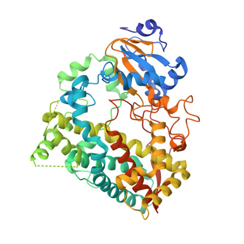
Structural Insights into the Interaction of Cytochrome P450 3A4 with Suicide Substrates: Mibefradil, Azamulin and 6',7'-Dihydroxybergamottin.

Sevrioukova, I.F.

(2019) Int J Mol Sci 20

  • DOI: https://doi.org/10.3390/ijms20174245
  • Primary Citation of Related Structures:  
    6OO9, 6OOA, 6OOB

  • PubMed Abstract: 

    Human cytochrome P450 3A4 (CYP3A4) is the most important drug-metabolizing enzyme. Some drugs and natural compounds can act as suicide (mechanism-based) inactivators of CYP3A4, leading to unanticipated drug-drug interactions, toxicity and therapeutic failures. Despite significant clinical and toxicological implications, the mechanism-based inactivation remains incompletely understood. This study provides the first direct insights into the interaction of CYP3A4 with three suicide substrates: mibefradil, an antihypertensive drug quickly withdrawn from the market; a semi-synthetic antibiotic azamulin; and a natural furanocoumarin, 6',7'-dihydroxybergamottin. Novel structural findings help better understand the suicide substrate binding and inhibitory mechanism, and can be used to improve the predictability of the binding ability, metabolic sites and inhibitory/inactivation potential of newly developed drugs and other chemicals relevant to public health.


  • Organizational Affiliation

    Department of Molecular Biology and Biochemistry, University of California, Irvine, CA 92697-3900, USA. sevrioui@uci.edu.


Macromolecules
Find similar proteins by:  (by identity cutoff)  |  3D Structure
Entity ID: 1
MoleculeChains Sequence LengthOrganismDetailsImage
Cytochrome P450 3A4487Homo sapiensMutation(s): 0 
Gene Names: CYP3A4CYP3A3
EC: 1.14.14 (PDB Primary Data), 1.14.14.56 (PDB Primary Data), 1.14.14.73 (PDB Primary Data), 1.14.14.55 (PDB Primary Data)
UniProt & NIH Common Fund Data Resources
Find proteins for P08684 (Homo sapiens)
Explore P08684 
Go to UniProtKB:  P08684
PHAROS:  P08684
GTEx:  ENSG00000160868 
Entity Groups  
Sequence Clusters30% Identity50% Identity70% Identity90% Identity95% Identity100% Identity
UniProt GroupP08684
Sequence Annotations
Expand
  • Reference Sequence
Small Molecules
Ligands 2 Unique
IDChains Name / Formula / InChI Key2D Diagram3D Interactions
HEM
Query on HEM

Download Ideal Coordinates CCD File 
B [auth A]PROTOPORPHYRIN IX CONTAINING FE
C34 H32 Fe N4 O4
KABFMIBPWCXCRK-RGGAHWMASA-L
MWS (Subject of Investigation/LOI)
Query on MWS

Download Ideal Coordinates CCD File 
C [auth A]4-{[(2Z,6S)-6,7-dihydroxy-3,7-dimethyloct-2-en-1-yl]oxy}-7H-furo[3,2-g][1]benzopyran-7-one
C21 H24 O6
IXZUPBUEKFXTSD-ZHJQRIIBSA-N
Experimental Data & Validation

Experimental Data

  • Method: X-RAY DIFFRACTION
  • Resolution: 2.20 Å
  • R-Value Free: 0.275 
  • R-Value Work: 0.207 
  • R-Value Observed: 0.210 
  • Space Group: I 2 2 2
Unit Cell:
Length ( Å )Angle ( ˚ )
a = 77.906α = 90
b = 102.383β = 90
c = 127.464γ = 90
Software Package:
Software NamePurpose
PHENIXrefinement
iMOSFLMdata reduction
SCALAdata scaling
PHASERphasing

Structure Validation

View Full Validation Report

Currently 6OOB does not have a validation slider image.


Entry History & Funding Information

Deposition Data


Funding OrganizationLocationGrant Number
National Institutes of Health/National Institute of Environmental Health Sciences (NIH/NIEHS)United StatesES025767

Revision History  (Full details and data files)

  • Version 1.0: 2019-09-11
    Type: Initial release
  • Version 1.1: 2019-09-18
    Changes: Data collection, Database references
  • Version 1.2: 2019-12-18
    Changes: Author supporting evidence
  • Version 1.3: 2023-10-11
    Changes: Data collection, Database references, Refinement description