6M5E

Human serum albumin with cyclic peptide dalbavancin


Experimental Data Snapshot

  • Method: X-RAY DIFFRACTION
  • Resolution: 2.80 Å
  • R-Value Free: 0.228 
  • R-Value Work: 0.180 
  • R-Value Observed: 0.182 

wwPDB Validation   3D Report Full Report


Ligand Structure Quality Assessment 


This is version 1.3 of the entry. See complete history


Literature

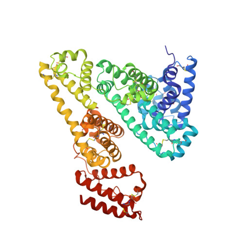
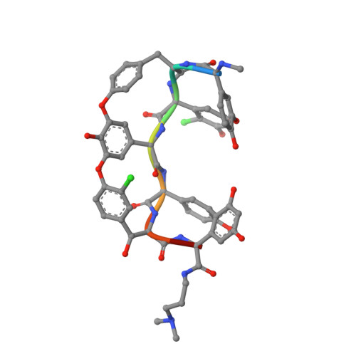
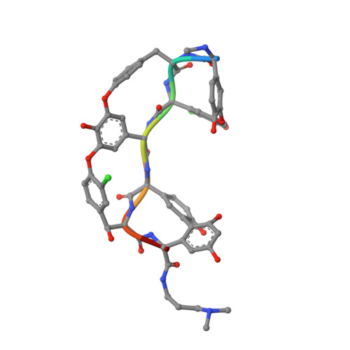
Structural Basis for the Binding Mechanism of Human Serum Albumin Complexed with Cyclic Peptide Dalbavancin.

Ito, S.Senoo, A.Nagatoishi, S.Ohue, M.Yamamoto, M.Tsumoto, K.Wakui, N.

(2020) J Med Chem 63: 14045-14053

  • DOI: https://doi.org/10.1021/acs.jmedchem.0c01578
  • Primary Citation of Related Structures:  
    6M5D, 6M5E

  • PubMed Abstract: 

    Cyclic peptides, with unique structural features, have emerged as new candidates for drug discovery; their association with human serum albumin (HSA; long blood half-life) is crucial to improve drug delivery and avoid renal clearance. Here, we present the crystal structure of HSA complexed with dalbavancin, a clinically used cyclic peptide. Small-angle X-ray scattering and isothermal titration calorimetry experiments showed that the HSA-dalbavancin complex exists in a monomeric state; dalbavancin is only bound to the subdomain IA of HSA in solution. Structural analysis and MD simulation revealed that the swing of Phe70 and movement of the helix near dalbavancin were necessary for binding. The flip of Leu251 promoted the formation of the binding pocket with an induced-fit mechanism; moreover, the movement of the loop region including Glu60 increased the number of noncovalent interactions with HSA. These findings may support the development of new cyclic peptides for clinical use, particularly the elucidation of their binding mechanism to HSA.


  • Organizational Affiliation

    ROD (Single Crystal Analysis) Group, Application Laboratories, Rigaku Corporation, 3-9-12 Matsubara-cho, Akishima, Tokyo 196-8666, Japan.


Macromolecules
Find similar proteins by:  (by identity cutoff)  |  3D Structure
Entity ID: 1
MoleculeChains Sequence LengthOrganismDetailsImage
Serum albumin
A, B, C
585Homo sapiensMutation(s): 0 
Gene Names: ALBGIG20GIG42PRO0903PRO1708PRO2044PRO2619PRO2675UNQ696/PRO1341
UniProt & NIH Common Fund Data Resources
Find proteins for P02768 (Homo sapiens)
Explore P02768 
Go to UniProtKB:  P02768
PHAROS:  P02768
GTEx:  ENSG00000163631 
Entity Groups  
Sequence Clusters30% Identity50% Identity70% Identity90% Identity95% Identity100% Identity
UniProt GroupP02768
Sequence Annotations
Expand
  • Reference Sequence

Find similar proteins by:  Sequence   |   3D Structure  

Entity ID: 2
MoleculeChains Sequence LengthOrganismDetailsImage
dalbavancinD [auth F],
E [auth G],
F [auth H]
8synthetic constructMutation(s): 0 
Sequence Annotations
Expand
  • Reference Sequence

Find similar proteins by:  Sequence   |   3D Structure  

Entity ID: 3
MoleculeChains Sequence LengthOrganismDetailsImage
dalbavancinG [auth I],
H [auth J]
8synthetic constructMutation(s): 0 
Sequence Annotations
Expand
  • Reference Sequence
Small Molecules
Ligands 6 Unique
IDChains Name / Formula / InChI Key2D Diagram3D Interactions
JEF
Query on JEF

Download Ideal Coordinates CCD File 
I [auth A]
J [auth A]
K [auth B]
L [auth B]
M [auth C]
I [auth A],
J [auth A],
K [auth B],
L [auth B],
M [auth C],
N [auth C]
O-(O-(2-AMINOPROPYL)-O'-(2-METHOXYETHYL)POLYPROPYLENE GLYCOL 500)
C30 H63 N O10
ICCXIDTYQFYPNV-RUMGZKRTSA-N
M12
Query on M12

Download Ideal Coordinates CCD File 
AA [auth J],
P [auth F],
S [auth G],
V [auth H],
X [auth I]
10-METHYLUNDECANOIC ACID
C12 H24 O2
QJRRBVNPIKYRQJ-UHFFFAOYSA-N
F8X
Query on F8X

Download Ideal Coordinates CCD File 
BA [auth J],
Y [auth I]
2-amino-2-deoxy-beta-D-altropyranuronic acid
C6 H11 N O6
CRIPFXSBMSGPKB-SYJWYVCOSA-N
F8F
Query on F8F

Download Ideal Coordinates CCD File 
O [auth F],
R [auth G],
U [auth H]
(2S,5S)-5-azanyl-3,4,6-tris(oxidanyl)oxane-2-carboxylic acid
C6 H11 N O6
CRIPFXSBMSGPKB-WAXACMCWSA-N
WOO
Query on WOO

Download Ideal Coordinates CCD File 
CA [auth J],
Z [auth I]
beta-L-allopyranose
C6 H12 O6
WQZGKKKJIJFFOK-RUTHBDMASA-N
MAN
Query on MAN

Download Ideal Coordinates CCD File 
Q [auth F],
T [auth G],
W [auth H]
alpha-D-mannopyranose
C6 H12 O6
WQZGKKKJIJFFOK-PQMKYFCFSA-N
Modified Residues  1 Unique
IDChains TypeFormula2D DiagramParent
5PG
Query on 5PG
D [auth F],
E [auth G],
F [auth H]
L-PEPTIDE LINKINGC9 H11 N O3GLY
Experimental Data & Validation

Experimental Data

  • Method: X-RAY DIFFRACTION
  • Resolution: 2.80 Å
  • R-Value Free: 0.228 
  • R-Value Work: 0.180 
  • R-Value Observed: 0.182 
  • Space Group: C 1 2 1
Unit Cell:
Length ( Å )Angle ( ˚ )
a = 197.05α = 90
b = 125.68β = 90.32
c = 125.74γ = 90
Software Package:
Software NamePurpose
PHENIXrefinement
XSCALEdata scaling
PHASERphasing
PDB_EXTRACTdata extraction
XDSdata reduction

Structure Validation

View Full Validation Report


Ligand Structure Quality Assessment 


Entry History & Funding Information

Deposition Data


Funding OrganizationLocationGrant Number
Japan Agency for Medical Research and Development (AMED)JapanJP19am0101001 (Support number 1851)

Revision History  (Full details and data files)

  • Version 1.0: 2020-11-18
    Type: Initial release
  • Version 1.1: 2020-12-02
    Changes: Database references
  • Version 1.2: 2020-12-09
    Changes: Database references
  • Version 1.3: 2023-11-29
    Changes: Data collection, Database references, Derived calculations, Refinement description