6LIO

Crystal structure of human PDK2 complexed with GM67520


Experimental Data Snapshot

  • Method: X-RAY DIFFRACTION
  • Resolution: 1.76 Å
  • R-Value Free: 0.269 
  • R-Value Work: 0.234 
  • R-Value Observed: 0.236 

wwPDB Validation   3D Report Full Report


Ligand Structure Quality Assessment 


This is version 1.1 of the entry. See complete history


Literature

Structural basis for the inhibition of PDK2 by novel ATP- and lipoyl-binding site targeting compounds.

Kang, J.Pagire, H.S.Kang, D.Song, Y.H.Lee, I.K.Lee, K.T.Park, C.J.Ahn, J.H.Kim, J.

(2020) Biochem Biophys Res Commun 527: 778-784

  • DOI: https://doi.org/10.1016/j.bbrc.2020.04.102
  • Primary Citation of Related Structures:  
    6LIL, 6LIN, 6LIO

  • PubMed Abstract: 

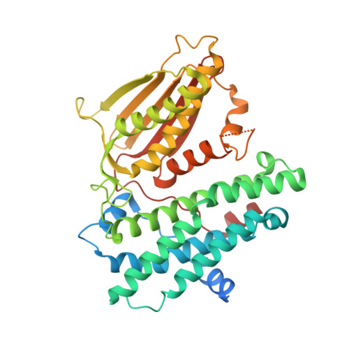
    Pyruvate dehydrogenase kinase (PDK) controls the activity of pyruvate decarboxylase complex (PDC) by phosphorylating key serine residues on the E1 subunit, which leads to a decreased oxidative phosphorylation in mitochondria. Inhibition of PDK activity by natural/synthetic compounds has been shown to reverse the Warburg effect, a characteristic metabolism in cancer cells. PDK-PDC axis also has been associated with diabetes and heart disease. Therefore, regulation of PDK activity has been considered as a promising strategy to treat related diseases. Here we present the X-ray crystal structure of PDK2 complexed with a recently identified PDK4 inhibitor, compound 8c, which has been predicted to bind at the lipoyl-binding site and interrupt intermolecular interactions with the E2-E3bp subunits of PDC. The co-crystal structure confirmed the specific binding location of compound 8c and revealed the remote conformational change in the ATP-binding pocket. In addition, two novel 4,5-diarylisoxazole derivatives, GM10030 and GM67520, were synthesized and used for structural studies, which target the ATP-binding site of PDK2. These compounds bind to PDK2 with a sub-100nM affinity as determined by isothermal titration calorimetry experiments. Notably, the crystal structure of the PDK2-GM10030 complex displays unprecedented asymmetric conformation of human PDK2 dimer, especially in the ATP-lids and C-terminal tails.


  • Organizational Affiliation

    Department of Chemistry, Gwangju Institute of Science and Technology, Gwangju, 61005, Republic of Korea.


Macromolecules
Find similar proteins by:  (by identity cutoff)  |  3D Structure
Entity ID: 1
MoleculeChains Sequence LengthOrganismDetailsImage
[Pyruvate dehydrogenase (acetyl-transferring)] kinase isozyme 2, mitochondrial
A, B
411Homo sapiensMutation(s): 0 
Gene Names: PDK2PDHK2
EC: 2.7.11.2
UniProt & NIH Common Fund Data Resources
Find proteins for Q15119 (Homo sapiens)
Explore Q15119 
Go to UniProtKB:  Q15119
PHAROS:  Q15119
GTEx:  ENSG00000005882 
Entity Groups  
Sequence Clusters30% Identity50% Identity70% Identity90% Identity95% Identity100% Identity
UniProt GroupQ15119
Sequence Annotations
Expand
  • Reference Sequence
Small Molecules
Ligands 3 Unique
IDChains Name / Formula / InChI Key2D Diagram3D Interactions
EH3 (Subject of Investigation/LOI)
Query on EH3

Download Ideal Coordinates CCD File 
G [auth A],
K [auth B]
4-[[[5-[5-chloranyl-2,4-bis(oxidanyl)phenyl]-4-[4-(1-methylsulfonylpiperidin-4-yl)oxyphenyl]-1,2-oxazol-3-yl]carbonylamino]methyl]cyclohexane-1-carboxylic acid
C30 H34 Cl N3 O9 S
AKZGGZLLGOJKMH-IZAXUBKRSA-N
SO4
Query on SO4

Download Ideal Coordinates CCD File 
D [auth A],
E [auth A],
F [auth A],
J [auth B]
SULFATE ION
O4 S
QAOWNCQODCNURD-UHFFFAOYSA-L
GOL
Query on GOL

Download Ideal Coordinates CCD File 
C [auth A],
H [auth B],
I [auth B]
GLYCEROL
C3 H8 O3
PEDCQBHIVMGVHV-UHFFFAOYSA-N
Binding Affinity Annotations 
IDSourceBinding Affinity
EH3 Binding MOAD:  6LIO Kd: 64.4 (nM) from 1 assay(s)
Experimental Data & Validation

Experimental Data

  • Method: X-RAY DIFFRACTION
  • Resolution: 1.76 Å
  • R-Value Free: 0.269 
  • R-Value Work: 0.234 
  • R-Value Observed: 0.236 
  • Space Group: P 21 21 21
Unit Cell:
Length ( Å )Angle ( ˚ )
a = 56.949α = 90
b = 95.122β = 90
c = 162.503γ = 90
Software Package:
Software NamePurpose
REFMACrefinement
HKL-2000data scaling
PDB_EXTRACTdata extraction
HKL-2000data reduction
MOLREPphasing

Structure Validation

View Full Validation Report


Ligand Structure Quality Assessment 


Entry History & Funding Information

Deposition Data


Funding OrganizationLocationGrant Number
National Research Foundation (Korea)Korea, Republic Of2016R1D1A1B03930716

Revision History  (Full details and data files)

  • Version 1.0: 2020-09-30
    Type: Initial release
  • Version 1.1: 2023-11-22
    Changes: Data collection, Database references, Refinement description