6LH4

Crystal structural of MacroD1-ADPr complex


Experimental Data Snapshot

  • Method: X-RAY DIFFRACTION
  • Resolution: 2.00 Å
  • R-Value Free: 0.285 
  • R-Value Work: 0.240 
  • R-Value Observed: 0.242 

wwPDB Validation   3D Report Full Report


Ligand Structure Quality Assessment 


This is version 1.3 of the entry. See complete history


Literature

Molecular basis for the MacroD1-mediated hydrolysis of ADP-ribosylation.

Yang, X.Ma, Y.Li, Y.Dong, Y.Yu, L.L.Wang, H.Guo, L.Wu, C.Yu, X.Liu, X.

(2020) DNA Repair (Amst) 94: 102899-102899

  • DOI: https://doi.org/10.1016/j.dnarep.2020.102899
  • Primary Citation of Related Structures:  
    6LH4

  • PubMed Abstract: 

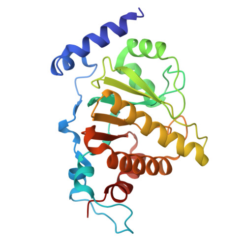
    MacroD1 is an enzyme that hydrolyzes protein mono-ADP-ribosylation. However, the key catalytic residues of MacroD1 in these biochemical reactions remain elusive. Here, we present the crystal structure of MacroD1 in a complex with ADP-ribose (ADPR). The β5-α10-loop functions as a switch loop to mediate substrate recognition and right orientation. The conserved Phe 272 in the β5-α10-loop plays a crucial role in the orientation of ADPR distal ribose, and a conserved hydrogen-bond network contributes significantly to hold and orient the catalytic water12, which mediates ADPR hydrolysis. Moreover, we found that MacroD1 was recruited to the sites of DNA damage via recognition of ADP-ribosylation at DNA lesions. The MacroD1-mediated ADPR hydrolysis is essential for DNA damage repair. Taken together, our study provides structural and functional insights into the molecular mechanism of MacroD1-mediated ADPR hydrolysis and its role in DNA damage repair.


  • Organizational Affiliation

    College of Life Science, Institute of Life Science and Green Development, Hebei University, Baoding, 071000, Hebei, PR China.


Macromolecules
Find similar proteins by:  (by identity cutoff)  |  3D Structure
Entity ID: 1
MoleculeChains Sequence LengthOrganismDetailsImage
ADP-ribose glycohydrolase MACROD1
A, B, C, D
235Homo sapiensMutation(s): 0 
Gene Names: MACROD1LRP16
EC: 3.1.1.106 (PDB Primary Data), 3.2.2 (PDB Primary Data)
UniProt & NIH Common Fund Data Resources
Find proteins for Q9BQ69 (Homo sapiens)
Explore Q9BQ69 
Go to UniProtKB:  Q9BQ69
PHAROS:  Q9BQ69
GTEx:  ENSG00000133315 
Entity Groups  
Sequence Clusters30% Identity50% Identity70% Identity90% Identity95% Identity100% Identity
UniProt GroupQ9BQ69
Sequence Annotations
Expand
  • Reference Sequence
Small Molecules
Ligands 1 Unique
IDChains Name / Formula / InChI Key2D Diagram3D Interactions
AR6 (Subject of Investigation/LOI)
Query on AR6

Download Ideal Coordinates CCD File 
E [auth A],
F [auth B],
G [auth C],
H [auth D]
[(2R,3S,4R,5R)-5-(6-AMINOPURIN-9-YL)-3,4-DIHYDROXY-OXOLAN-2-YL]METHYL [HYDROXY-[[(2R,3S,4R,5S)-3,4,5-TRIHYDROXYOXOLAN-2-YL]METHOXY]PHOSPHORYL] HYDROGEN PHOSPHATE
C15 H23 N5 O14 P2
SRNWOUGRCWSEMX-ZQSHOCFMSA-N
Experimental Data & Validation

Experimental Data

  • Method: X-RAY DIFFRACTION
  • Resolution: 2.00 Å
  • R-Value Free: 0.285 
  • R-Value Work: 0.240 
  • R-Value Observed: 0.242 
  • Space Group: P 1 21 1
Unit Cell:
Length ( Å )Angle ( ˚ )
a = 79.9α = 90
b = 106.2β = 120
c = 79.9γ = 90
Software Package:
Software NamePurpose
PHENIXrefinement
XDSdata reduction
XSCALEdata scaling
PHASERphasing
PDB_EXTRACTdata extraction

Structure Validation

View Full Validation Report


Ligand Structure Quality Assessment 


Entry History & Funding Information

Deposition Data


Funding OrganizationLocationGrant Number
National Natural Science Foundation of China (NSFC)China81672794

Revision History  (Full details and data files)

  • Version 1.0: 2020-12-09
    Type: Initial release
  • Version 1.1: 2021-06-23
    Changes: Database references
  • Version 1.2: 2023-02-15
    Changes: Advisory, Database references, Derived calculations
  • Version 1.3: 2023-11-29
    Changes: Data collection, Refinement description