6KDY

Crystal structure of the alpha bata heterodimer of human IDH3 in complex with NAD.


Experimental Data Snapshot

  • Method: X-RAY DIFFRACTION
  • Resolution: 3.02 Å
  • R-Value Free: 0.246 
  • R-Value Work: 0.197 
  • R-Value Observed: 0.200 

wwPDB Validation   3D Report Full Report


Ligand Structure Quality Assessment 


This is version 1.2 of the entry. See complete history


Literature

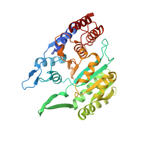
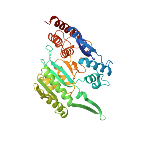
Molecular basis for the function of the alpha beta heterodimer of human NAD-dependent isocitrate dehydrogenase.

Sun, P.Ma, T.Zhang, T.Zhu, H.Zhang, J.Liu, Y.Ding, J.

(2019) J Biol Chem 294: 16214-16227

  • DOI: https://doi.org/10.1074/jbc.RA119.010099
  • Primary Citation of Related Structures:  
    6KDE, 6KDF, 6KDY, 6KE3

  • PubMed Abstract: 

    Mammalian mitochondrial NAD-dependent isocitrate dehydrogenase (NAD-IDH) catalyzes the decarboxylation of isocitrate into α-ketoglutarate in the tricarboxylic acid cycle. It exists as the α 2 βγ heterotetramer composed of the αβ and αγ heterodimers. Different from the αγ heterodimer that can be allosterically activated by CIT and ADP, the αβ heterodimer cannot be allosterically regulated by the activators; however, the molecular mechanism is unclear. We report here the crystal structures of the αβ heterodimer of human NAD-IDH with the α subunit in apo form and in Ca 2+ -bound, NAD-bound, and NADH-bound forms. Structural analyses and comparisons reveal that the αβ heterodimer has a similar yet more compact overall structure compared with the αγ heterodimer and contains a pseudo-allosteric site that is structurally different from the allosteric site. In particular, the β3-α3 and β12-α8 loops of the β subunit at the pseudo-allosteric site adopt significantly different conformations from those of the γ subunit at the allosteric site and hence impede the binding of the activators, explaining why the αβ heterodimer cannot be allosterically regulated by the activators. The structural data also show that NADH can compete with NAD to bind to the active site and inhibits the activity of the αβ heterodimer. These findings together with the biochemical data reveal the molecular basis for the function of the αβ heterodimer of human NAD-IDH.


  • Organizational Affiliation

    State Key Laboratory of Molecular Biology, CAS Center for Excellence in Molecular Cell Science, Institute of Biochemistry and Cell Biology, University of Chinese Academy of Sciences, Chinese Academy of Sciences, 320 Yue-Yang Road, Shanghai 200031, China.


Macromolecules
Find similar proteins by:  (by identity cutoff)  |  3D Structure
Entity ID: 1
MoleculeChains Sequence LengthOrganismDetailsImage
Isocitrate dehydrogenase [NAD] subunit alpha, mitochondrial
A, C, E, G
341Homo sapiensMutation(s): 0 
Gene Names: IDH3A
EC: 1.1.1.41
UniProt & NIH Common Fund Data Resources
Find proteins for P50213 (Homo sapiens)
Explore P50213 
Go to UniProtKB:  P50213
PHAROS:  P50213
GTEx:  ENSG00000166411 
Entity Groups  
Sequence Clusters30% Identity50% Identity70% Identity90% Identity95% Identity100% Identity
UniProt GroupP50213
Sequence Annotations
Expand
  • Reference Sequence
Find similar proteins by:  (by identity cutoff)  |  3D Structure
Entity ID: 2
MoleculeChains Sequence LengthOrganismDetailsImage
Isocitrate dehydrogenase [NAD] subunit beta, mitochondrial
B, D, F, H
356Homo sapiensMutation(s): 0 
Gene Names: IDH3B
UniProt & NIH Common Fund Data Resources
Find proteins for O43837 (Homo sapiens)
Explore O43837 
Go to UniProtKB:  O43837
PHAROS:  O43837
GTEx:  ENSG00000101365 
Entity Groups  
Sequence Clusters30% Identity50% Identity70% Identity90% Identity95% Identity100% Identity
UniProt GroupO43837
Sequence Annotations
Expand
  • Reference Sequence
Small Molecules
Ligands 5 Unique
IDChains Name / Formula / InChI Key2D Diagram3D Interactions
NAD (Subject of Investigation/LOI)
Query on NAD

Download Ideal Coordinates CCD File 
I [auth A],
K [auth C],
N [auth E],
P [auth G]
NICOTINAMIDE-ADENINE-DINUCLEOTIDE
C21 H27 N7 O14 P2
BAWFJGJZGIEFAR-NNYOXOHSSA-N
2PE
Query on 2PE

Download Ideal Coordinates CCD File 
L [auth D]NONAETHYLENE GLYCOL
C18 H38 O10
YZUUTMGDONTGTN-UHFFFAOYSA-N
PE7
Query on PE7

Download Ideal Coordinates CCD File 
O [auth F]1-DEOXY-1-THIO-HEPTAETHYLENE GLYCOL
C14 H30 O7 S
ACMBXVJDKVNCGH-UHFFFAOYSA-N
P6G
Query on P6G

Download Ideal Coordinates CCD File 
Q [auth H]HEXAETHYLENE GLYCOL
C12 H26 O7
IIRDTKBZINWQAW-UHFFFAOYSA-N
PG4
Query on PG4

Download Ideal Coordinates CCD File 
J [auth B],
M [auth D]
TETRAETHYLENE GLYCOL
C8 H18 O5
UWHCKJMYHZGTIT-UHFFFAOYSA-N
Experimental Data & Validation

Experimental Data

  • Method: X-RAY DIFFRACTION
  • Resolution: 3.02 Å
  • R-Value Free: 0.246 
  • R-Value Work: 0.197 
  • R-Value Observed: 0.200 
  • Space Group: P 1 21 1
Unit Cell:
Length ( Å )Angle ( ˚ )
a = 99.46α = 90
b = 162.979β = 100.31
c = 114.407γ = 90
Software Package:
Software NamePurpose
PHENIXrefinement
HKL-2000data scaling
PDB_EXTRACTdata extraction
HKL-2000data reduction
PHASERphasing

Structure Validation

View Full Validation Report


Ligand Structure Quality Assessment 


Entry History & Funding Information

Deposition Data


Funding OrganizationLocationGrant Number
National Natural Science Foundation of ChinaChina--

Revision History  (Full details and data files)

  • Version 1.0: 2019-09-25
    Type: Initial release
  • Version 1.1: 2019-12-25
    Changes: Database references, Structure summary
  • Version 1.2: 2023-11-22
    Changes: Data collection, Database references, Refinement description