6K0R

Ruvbl1-Ruvbl2 with truncated domain II in complex with phosphorylated Cordycepin


Experimental Data Snapshot

  • Method: X-RAY DIFFRACTION
  • Resolution: 2.50 Å
  • R-Value Free: 0.269 
  • R-Value Work: 0.228 
  • R-Value Observed: 0.230 

wwPDB Validation   3D Report Full Report


Ligand Structure Quality Assessment 


This is version 1.2 of the entry. See complete history


Literature

Chemical perturbations reveal that RUVBL2 regulates the circadian phase in mammals.

Ju, D.Zhang, W.Yan, J.Zhao, H.Li, W.Wang, J.Liao, M.Xu, Z.Wang, Z.Zhou, G.Mei, L.Hou, N.Ying, S.Cai, T.Chen, S.Xie, X.Lai, L.Tang, C.Park, N.Takahashi, J.S.Huang, N.Qi, X.Zhang, E.E.

(2020) Sci Transl Med 12

  • DOI: https://doi.org/10.1126/scitranslmed.aba0769
  • Primary Citation of Related Structures:  
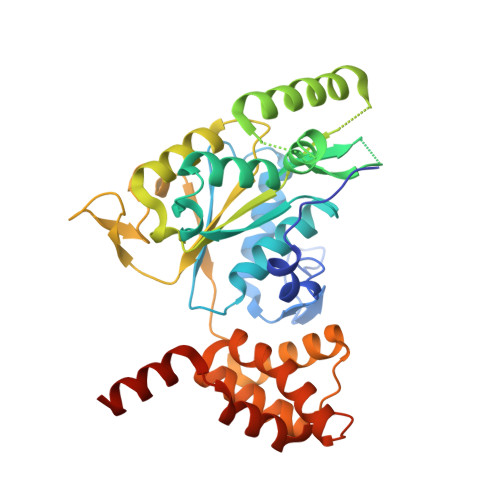
    6K0R

  • PubMed Abstract: 

    Transcriptional regulation lies at the core of the circadian clockwork, but how the clock-related transcription machinery controls the circadian phase is not understood. Here, we show both in human cells and in mice that RuvB-like ATPase 2 (RUVBL2) interacts with other known clock proteins on chromatin to regulate the circadian phase. Pharmacological perturbation of RUVBL2 with the adenosine analog compound cordycepin resulted in a rapid-onset 12-hour clock phase-shift phenotype at human cell, mouse tissue, and whole-animal live imaging levels. Using simple peripheral injection treatment, we found that cordycepin penetrated the blood-brain barrier and caused rapid entrainment of the circadian phase, facilitating reduced duration of recovery in a mouse jet-lag model. We solved a crystal structure for human RUVBL2 in complex with a physiological metabolite of cordycepin, and biochemical assays showed that cordycepin treatment caused disassembly of an interaction between RUVBL2 and the core clock component BMAL1. Moreover, we showed with spike-in ChIP-seq analysis and binding assays that cordycepin treatment caused disassembly of the circadian super-complex, which normally resides at E-box chromatin loci such as PER1 , PER2 , DBP, and NR1D1 Mathematical modeling supported that the observed type 0 phase shifts resulted from derepression of E-box clock gene transcription.


  • Organizational Affiliation

    National Institute of Biological Sciences, Beijing 102206, China.


Macromolecules
Find similar proteins by:  (by identity cutoff)  |  3D Structure
Entity ID: 1
MoleculeChains Sequence LengthOrganismDetailsImage
RuvB-like 1,RuvB-like 1
A, B, C, G, H
A, B, C, G, H, I
355Homo sapiensMutation(s): 0 
Gene Names: RUVBL1INO80HNMP238TIP49TIP49A
EC: 3.6.4.12
UniProt & NIH Common Fund Data Resources
Find proteins for Q9Y265 (Homo sapiens)
Explore Q9Y265 
Go to UniProtKB:  Q9Y265
PHAROS:  Q9Y265
GTEx:  ENSG00000175792 
Entity Groups  
Sequence Clusters30% Identity50% Identity70% Identity90% Identity95% Identity100% Identity
UniProt GroupQ9Y265
Sequence Annotations
Expand
  • Reference Sequence
Find similar proteins by:  (by identity cutoff)  |  3D Structure
Entity ID: 2
MoleculeChains Sequence LengthOrganismDetailsImage
RuvB-like 2,RuvB-like 2
D, E, F, J, K
D, E, F, J, K, L
366Homo sapiensMutation(s): 0 
Gene Names: RUVBL2INO80JTIP48TIP49BCGI-46
EC: 3.6.4.12
UniProt & NIH Common Fund Data Resources
Find proteins for Q9Y230 (Homo sapiens)
Explore Q9Y230 
Go to UniProtKB:  Q9Y230
PHAROS:  Q9Y230
GTEx:  ENSG00000183207 
Entity Groups  
Sequence Clusters30% Identity50% Identity70% Identity90% Identity95% Identity100% Identity
UniProt GroupQ9Y230
Sequence Annotations
Expand
  • Reference Sequence
Small Molecules
Ligands 5 Unique
IDChains Name / Formula / InChI Key2D Diagram3D Interactions
3AT (Subject of Investigation/LOI)
Query on 3AT

Download Ideal Coordinates CCD File 
R [auth E],
W [auth J]
3'-DEOXYADENOSINE-5'-TRIPHOSPHATE
C10 H16 N5 O12 P3
NLIHPCYXRYQPSD-BAJZRUMYSA-N
ADP
Query on ADP

Download Ideal Coordinates CCD File 
M [auth A]ADENOSINE-5'-DIPHOSPHATE
C10 H15 N5 O10 P2
XTWYTFMLZFPYCI-KQYNXXCUSA-N
CUU (Subject of Investigation/LOI)
Query on CUU

Download Ideal Coordinates CCD File 
N [auth B]
O [auth C]
P [auth D]
S [auth F]
T [auth G]
N [auth B],
O [auth C],
P [auth D],
S [auth F],
T [auth G],
U [auth H],
V [auth I],
X [auth K]
[(2~{S},4~{R},5~{R})-5-(6-aminopurin-9-yl)-4-oxidanyl-oxolan-2-yl]methyl phosphono hydrogen phosphate
C10 H15 N5 O9 P2
OBEBFUHFXYDSAW-BAJZRUMYSA-N
CU0
Query on CU0

Download Ideal Coordinates CCD File 
Y [auth L][(2~{R},3~{S},4~{S})-3,4-bis(oxidanyl)oxolan-2-yl]methyl phosphono hydrogen phosphate
C5 H12 O10 P2
HNPYBPXXPMVWIY-LMVFSUKVSA-N
MG (Subject of Investigation/LOI)
Query on MG

Download Ideal Coordinates CCD File 
Q [auth D],
Z [auth L]
MAGNESIUM ION
Mg
JLVVSXFLKOJNIY-UHFFFAOYSA-N
Experimental Data & Validation

Experimental Data

  • Method: X-RAY DIFFRACTION
  • Resolution: 2.50 Å
  • R-Value Free: 0.269 
  • R-Value Work: 0.228 
  • R-Value Observed: 0.230 
  • Space Group: C 1 2 1
Unit Cell:
Length ( Å )Angle ( ˚ )
a = 110.304α = 90
b = 186.424β = 91.04
c = 235.176γ = 90
Software Package:
Software NamePurpose
BUSTERrefinement
PDB_EXTRACTdata extraction
XDSdata reduction
STARANISOdata scaling
PHASERphasing

Structure Validation

View Full Validation Report


Ligand Structure Quality Assessment 


Entry History 

Deposition Data

Revision History  (Full details and data files)

  • Version 1.0: 2020-05-06
    Type: Initial release
  • Version 1.1: 2020-05-20
    Changes: Database references
  • Version 1.2: 2023-11-22
    Changes: Data collection, Database references, Refinement description