6J9J

crystal structure of SESTD2 in complex with H3.3S31phK36M peptide


Experimental Data Snapshot

  • Method: X-RAY DIFFRACTION
  • Resolution: 1.78 Å
  • R-Value Free: 0.206 
  • R-Value Work: 0.189 
  • R-Value Observed: 0.190 

wwPDB Validation   3D Report Full Report


Ligand Structure Quality Assessment 


This is version 1.2 of the entry. See complete history


Literature

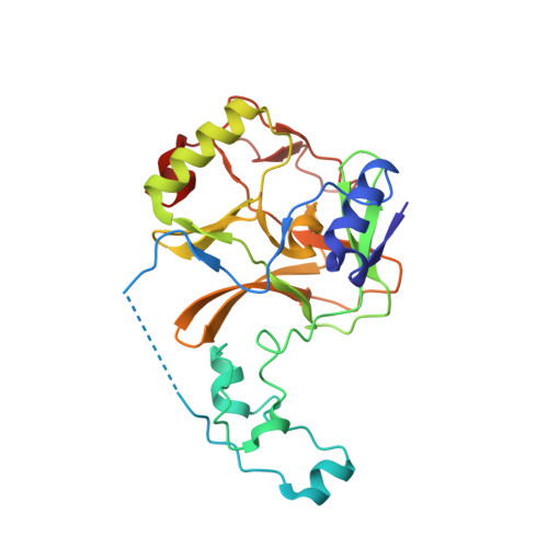
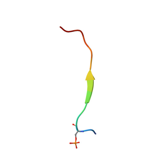
Histone H3.3 phosphorylation amplifies stimulation-induced transcription.

Armache, A.Yang, S.Martinez de Paz, A.Robbins, L.E.Durmaz, C.Cheong, J.Q.Ravishankar, A.Daman, A.W.Ahimovic, D.J.Klevorn, T.Yue, Y.Arslan, T.Lin, S.Panchenko, T.Hrit, J.Wang, M.Thudium, S.Garcia, B.A.Korb, E.Armache, K.J.Rothbart, S.B.Hake, S.B.Allis, C.D.Li, H.Josefowicz, S.Z.

(2020) Nature 583: 852-857

  • DOI: https://doi.org/10.1038/s41586-020-2533-0
  • Primary Citation of Related Structures:  
    6J9J

  • PubMed Abstract: 

    Complex organisms can rapidly induce select genes in response to diverse environmental cues. This regulation occurs in the context of large genomes condensed by histone proteins into chromatin. The sensing of pathogens by macrophages engages conserved signalling pathways and transcription factors to coordinate the induction of inflammatory genes 1-3 . Enriched integration of histone H3.3, the ancestral histone H3 variant, is a general feature of dynamically regulated chromatin and transcription 4-7 . However, how chromatin is regulated at induced genes, and what features of H3.3 might enable rapid and high-level transcription, are unknown. The amino terminus of H3.3 contains a unique serine residue (Ser31) that is absent in 'canonical' H3.1 and H3.2. Here we show that this residue, H3.3S31, is phosphorylated (H3.3S31ph) in a stimulation-dependent manner along rapidly induced genes in mouse macrophages. This selective mark of stimulation-responsive genes directly engages the histone methyltransferase SETD2, a component of the active transcription machinery, and 'ejects' the elongation corepressor ZMYND11 8,9 . We propose that features of H3.3 at stimulation-induced genes, including H3.3S31ph, provide preferential access to the transcription apparatus. Our results indicate dedicated mechanisms that enable rapid transcription involving the histone variant H3.3, its phosphorylation, and both the recruitment and the ejection of chromatin regulators.


  • Organizational Affiliation

    Laboratory of Epigenetics and Immunity, Department of Pathology and Laboratory Medicine, Weill Cornell Medicine, New York, NY, USA.


Macromolecules
Find similar proteins by:  (by identity cutoff)  |  3D Structure
Entity ID: 1
MoleculeChains Sequence LengthOrganismDetailsImage
Histone-lysine N-methyltransferase SETD2257Homo sapiensMutation(s): 0 
Gene Names: SETD2HIF1HYPBKIAA1732KMT3ASET2HSPC069
EC: 2.1.1.43 (PDB Primary Data), 2.1.1 (PDB Primary Data)
UniProt & NIH Common Fund Data Resources
Find proteins for Q9BYW2 (Homo sapiens)
Explore Q9BYW2 
Go to UniProtKB:  Q9BYW2
PHAROS:  Q9BYW2
GTEx:  ENSG00000181555 
Entity Groups  
Sequence Clusters30% Identity50% Identity70% Identity90% Identity95% Identity100% Identity
UniProt GroupQ9BYW2
Sequence Annotations
Expand
  • Reference Sequence

Find similar proteins by:  Sequence   |   3D Structure  

Entity ID: 2
MoleculeChains Sequence LengthOrganismDetailsImage
H3.3S31phK36M(29-42)14Homo sapiensMutation(s): 0 
UniProt & NIH Common Fund Data Resources
Find proteins for P84243 (Homo sapiens)
Explore P84243 
Go to UniProtKB:  P84243
PHAROS:  P84243
Entity Groups  
Sequence Clusters30% Identity50% Identity70% Identity90% Identity95% Identity100% Identity
UniProt GroupP84243
Sequence Annotations
Expand
  • Reference Sequence
Small Molecules
Ligands 3 Unique
IDChains Name / Formula / InChI Key2D Diagram3D Interactions
SAH
Query on SAH

Download Ideal Coordinates CCD File 
F [auth A]S-ADENOSYL-L-HOMOCYSTEINE
C14 H20 N6 O5 S
ZJUKTBDSGOFHSH-WFMPWKQPSA-N
ZN
Query on ZN

Download Ideal Coordinates CCD File 
C [auth A],
D [auth A],
E [auth A]
ZINC ION
Zn
PTFCDOFLOPIGGS-UHFFFAOYSA-N
SCN
Query on SCN

Download Ideal Coordinates CCD File 
G [auth A],
H [auth A],
I [auth A],
J [auth A]
THIOCYANATE ION
C N S
ZMZDMBWJUHKJPS-UHFFFAOYSA-M
Modified Residues  1 Unique
IDChains TypeFormula2D DiagramParent
SEP
Query on SEP
B
L-PEPTIDE LINKINGC3 H8 N O6 PSER
Experimental Data & Validation

Experimental Data

  • Method: X-RAY DIFFRACTION
  • Resolution: 1.78 Å
  • R-Value Free: 0.206 
  • R-Value Work: 0.189 
  • R-Value Observed: 0.190 
  • Space Group: P 21 21 21
Unit Cell:
Length ( Å )Angle ( ˚ )
a = 61.112α = 90
b = 77.014β = 90
c = 77.412γ = 90
Software Package:
Software NamePurpose
REFMACrefinement
SCALEPACKdata scaling
MOLREPphasing
PDB_EXTRACTdata extraction
HKL-2000data reduction

Structure Validation

View Full Validation Report


Ligand Structure Quality Assessment 


Entry History 

Deposition Data

Revision History  (Full details and data files)

  • Version 1.0: 2020-02-26
    Type: Initial release
  • Version 1.1: 2020-08-05
    Changes: Database references, Derived calculations
  • Version 1.2: 2020-08-12
    Changes: Database references