6GKG

Structure of 14-3-3 gamma in complex with caspase-2 14-3-3 binding motif Ser164


Experimental Data Snapshot

  • Method: X-RAY DIFFRACTION
  • Resolution: 2.85 Å
  • R-Value Free: 0.287 
  • R-Value Work: 0.231 
  • R-Value Observed: 0.233 

wwPDB Validation   3D Report Full Report


This is version 1.2 of the entry. See complete history


Literature

14-3-3 protein masks the nuclear localization sequence of caspase-2.

Smidova, A.Alblova, M.Kalabova, D.Psenakova, K.Rosulek, M.Herman, P.Obsil, T.Obsilova, V.

(2018) FEBS J 285: 4196-4213

  • DOI: https://doi.org/10.1111/febs.14670
  • Primary Citation of Related Structures:  
    6GKF, 6GKG

  • PubMed Abstract: 

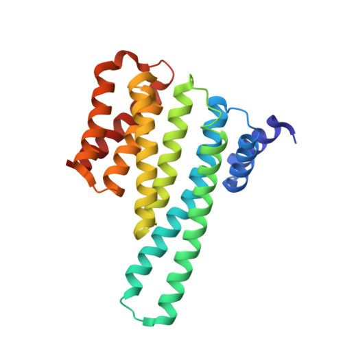
    Caspase-2 is an apical protease responsible for the proteolysis of cellular substrates directly involved in mediating apoptotic signaling cascades. Caspase-2 activation is inhibited by phosphorylation followed by binding to the scaffolding protein 14-3-3, which recognizes two phosphoserines located in the linker between the caspase recruitment domain and the p19 domains of the caspase-2 zymogen. However, the structural details of this interaction and the exact role of 14-3-3 in the regulation of caspase-2 activation remain unclear. Moreover, the caspase-2 region with both 14-3-3-binding motifs also contains the nuclear localization sequence (NLS), thus suggesting that 14-3-3 binding may regulate the subcellular localization of caspase-2. Here, we report a structural analysis of the 14-3-3ζ:caspase-2 complex using a combined approach based on small angle X-ray scattering, NMR, chemical cross-linking, and fluorescence spectroscopy. The structural model proposed in this study suggests that phosphorylated caspase-2 and 14-3-3ζ form a compact and rigid complex in which the p19 and the p12 domains of caspase-2 are positioned within the central channel of the 14-3-3 dimer and stabilized through interactions with the C-terminal helices of both 14-3-3ζ protomers. In this conformation, the surface of the p12 domain, which is involved in caspase-2 activation by dimerization, is sterically occluded by the 14-3-3 dimer, thereby likely preventing caspase-2 activation. In addition, 14-3-3 protein binding to caspase-2 masks its NLS. Therefore, our results suggest that 14-3-3 protein binding to caspase-2 may play a key role in regulating caspase-2 activation. DATABASE: The atomic coordinates and structure factors have been deposited in the Protein Data Bank, www.ww pdb.org (PDB ID codes 6GKF and 6GKG).


  • Organizational Affiliation

    Department of Structural Biology of Signaling Proteins, Division BIOCEV, Institute of Physiology of the Czech Academy of Sciences, Vestec, Czech Republic.


Macromolecules
Find similar proteins by:  (by identity cutoff)  |  3D Structure
Entity ID: 1
MoleculeChains Sequence LengthOrganismDetailsImage
14-3-3 protein gamma
A, B, C, D, E
A, B, C, D, E, F, G, H
234Homo sapiensMutation(s): 0 
Gene Names: YWHAG
UniProt & NIH Common Fund Data Resources
Find proteins for P61981 (Homo sapiens)
Explore P61981 
Go to UniProtKB:  P61981
PHAROS:  P61981
GTEx:  ENSG00000170027 
Entity Groups  
Sequence Clusters30% Identity50% Identity70% Identity90% Identity95% Identity100% Identity
UniProt GroupP61981
Sequence Annotations
Expand
  • Reference Sequence

Find similar proteins by:  Sequence   |   3D Structure  

Entity ID: 2
MoleculeChains Sequence LengthOrganismDetailsImage
Caspase-2
I, J, K, L, M
I, J, K, L, M, N
8Homo sapiensMutation(s): 0 
EC: 3.4.22.55
UniProt & NIH Common Fund Data Resources
Find proteins for P42575 (Homo sapiens)
Explore P42575 
Go to UniProtKB:  P42575
PHAROS:  P42575
GTEx:  ENSG00000106144 
Entity Groups  
UniProt GroupP42575
Sequence Annotations
Expand
  • Reference Sequence
Small Molecules
Modified Residues  1 Unique
IDChains TypeFormula2D DiagramParent
SEP
Query on SEP
I, J, K, L, M
I, J, K, L, M, N
L-PEPTIDE LINKINGC3 H8 N O6 PSER
Experimental Data & Validation

Experimental Data

  • Method: X-RAY DIFFRACTION
  • Resolution: 2.85 Å
  • R-Value Free: 0.287 
  • R-Value Work: 0.231 
  • R-Value Observed: 0.233 
  • Space Group: P 41 21 2
Unit Cell:
Length ( Å )Angle ( ˚ )
a = 121.987α = 90
b = 121.987β = 90
c = 311.004γ = 90
Software Package:
Software NamePurpose
PHENIXrefinement
XDSdata reduction
XDSdata scaling
MOLREPphasing

Structure Validation

View Full Validation Report


Entry History & Funding Information

Deposition Data


Funding OrganizationLocationGrant Number
Czech Science FoundationCzech Republic17-00726S

Revision History  (Full details and data files)

  • Version 1.0: 2018-10-17
    Type: Initial release
  • Version 1.1: 2018-11-28
    Changes: Data collection, Database references, Structure summary
  • Version 1.2: 2024-01-17
    Changes: Data collection, Database references, Refinement description