6GFV

M tuberculosis LpqI


Experimental Data Snapshot

  • Method: X-RAY DIFFRACTION
  • Resolution: 1.96 Å
  • R-Value Free: 0.268 
  • R-Value Work: 0.217 
  • R-Value Observed: 0.219 

wwPDB Validation   3D Report Full Report


This is version 1.3 of the entry. See complete history


Literature

The hydrolase LpqI primes mycobacterial peptidoglycan recycling.

Moynihan, P.J.Cadby, I.T.Veerapen, N.Jankute, M.Crosatti, M.Mukamolova, G.V.Lovering, A.L.Besra, G.S.

(2019) Nat Commun 10: 2647-2647

  • DOI: https://doi.org/10.1038/s41467-019-10586-2
  • Primary Citation of Related Structures:  
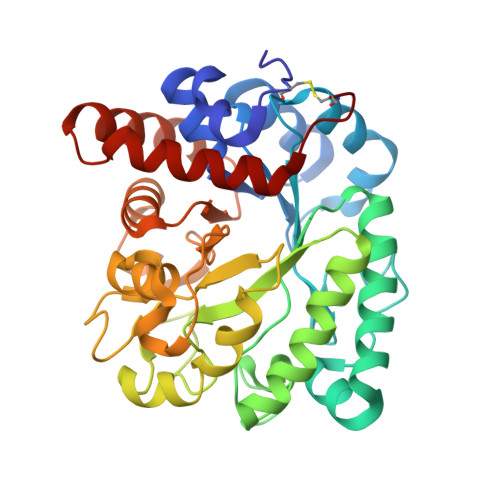
    6GFV

  • PubMed Abstract: 

    Growth and division by most bacteria requires remodelling and cleavage of their cell wall. A byproduct of this process is the generation of free peptidoglycan (PG) fragments known as muropeptides, which are recycled in many model organisms. Bacteria and hosts can harness the unique nature of muropeptides as a signal for cell wall damage and infection, respectively. Despite this critical role for muropeptides, it has long been thought that pathogenic mycobacteria such as Mycobacterium tuberculosis do not recycle their PG. Herein we show that M. tuberculosis and Mycobacterium bovis BCG are able to recycle components of their PG. We demonstrate that the core mycobacterial gene lpqI, encodes an authentic NagZ β-N-acetylglucosaminidase and that it is essential for PG-derived amino sugar recycling via an unusual pathway. Together these data provide a critical first step in understanding how mycobacteria recycle their peptidoglycan.


  • Organizational Affiliation

    Institute of Microbiology and Infection, School of Biological Sciences, University of Birmingham, Birmingham, B15 2TT, UK. p.j.moynihan@bham.ac.uk.


Macromolecules
Find similar proteins by:  (by identity cutoff)  |  3D Structure
Entity ID: 1
MoleculeChains Sequence LengthOrganismDetailsImage
Probable conserved lipoprotein LpqI
A, B
343Mycobacterium tuberculosis H37RvMutation(s): 0 
Gene Names: lpqIRv0237
UniProt
Find proteins for L7N6B0 (Mycobacterium tuberculosis (strain ATCC 25618 / H37Rv))
Explore L7N6B0 
Go to UniProtKB:  L7N6B0
Entity Groups  
Sequence Clusters30% Identity50% Identity70% Identity90% Identity95% Identity100% Identity
UniProt GroupL7N6B0
Sequence Annotations
Expand
  • Reference Sequence
Experimental Data & Validation

Experimental Data

  • Method: X-RAY DIFFRACTION
  • Resolution: 1.96 Å
  • R-Value Free: 0.268 
  • R-Value Work: 0.217 
  • R-Value Observed: 0.219 
  • Space Group: C 1 2 1
Unit Cell:
Length ( Å )Angle ( ˚ )
a = 121.821α = 90
b = 89.579β = 125.54
c = 87.107γ = 90
Software Package:
Software NamePurpose
XDSdata reduction
Aimlessdata scaling
PHASERphasing
PHENIXrefinement
PDB_EXTRACTdata extraction

Structure Validation

View Full Validation Report


Entry History & Funding Information

Deposition Data


Funding OrganizationLocationGrant Number
Biotechnology and Biological Sciences Research CouncilUnited KingdomBB/N011945/1

Revision History  (Full details and data files)

  • Version 1.0: 2019-05-15
    Type: Initial release
  • Version 1.1: 2019-05-29
    Changes: Data collection, Database references
  • Version 1.2: 2019-06-26
    Changes: Data collection, Database references
  • Version 1.3: 2024-01-17
    Changes: Data collection, Database references, Refinement description