6FY5

Crystal structure of the macro domain of human macroh2a2


Experimental Data Snapshot

  • Method: X-RAY DIFFRACTION
  • Resolution: 1.65 Å
  • R-Value Free: 0.244 
  • R-Value Work: 0.215 
  • R-Value Observed: 0.216 

wwPDB Validation   3D Report Full Report


This is version 1.3 of the entry. See complete history


Literature

MacroH2A histone variants limit chromatin plasticity through two distinct mechanisms.

Kozlowski, M.Corujo, D.Hothorn, M.Guberovic, I.Mandemaker, I.K.Blessing, C.Sporn, J.Gutierrez-Triana, A.Smith, R.Portmann, T.Treier, M.Scheffzek, K.Huet, S.Timinszky, G.Buschbeck, M.Ladurner, A.G.

(2018) EMBO Rep 19

  • DOI: https://doi.org/10.15252/embr.201744445
  • Primary Citation of Related Structures:  
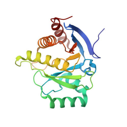
    6FY5

  • PubMed Abstract: 

    MacroH2A histone variants suppress tumor progression and act as epigenetic barriers to induced pluripotency. How they impart their influence on chromatin plasticity is not well understood. Here, we analyze how the different domains of macroH2A proteins contribute to chromatin structure and dynamics. By solving the crystal structure of the macrodomain of human macroH2A2 at 1.7 Å, we find that its putative binding pocket exhibits marked structural differences compared with the macroH2A1.1 isoform, rendering macroH2A2 unable to bind ADP-ribose. Quantitative binding assays show that this specificity is conserved among vertebrate macroH2A isoforms. We further find that macroH2A histones reduce the transient, PARP1-dependent chromatin relaxation that occurs in living cells upon DNA damage through two distinct mechanisms. First, macroH2A1.1 mediates an isoform-specific effect through its ability to suppress PARP1 activity. Second, the unstructured linker region exerts an additional repressive effect that is common to all macroH2A proteins. In the absence of DNA damage, the macroH2A linker is also sufficient for rescuing heterochromatin architecture in cells deficient for macroH2A.


  • Organizational Affiliation

    Biomedical Center, Physiological Chemistry, Ludwig-Maximilians-Universität München, Planegg-Martinsried, Germany.


Macromolecules
Find similar proteins by:  (by identity cutoff)  |  3D Structure
Entity ID: 1
MoleculeChains Sequence LengthOrganismDetailsImage
Core histone macro-H2A.2
A, B
198Homo sapiensMutation(s): 0 
Gene Names: H2AFY2MACROH2A2
UniProt & NIH Common Fund Data Resources
Find proteins for Q9P0M6 (Homo sapiens)
Explore Q9P0M6 
Go to UniProtKB:  Q9P0M6
PHAROS:  Q9P0M6
GTEx:  ENSG00000099284 
Entity Groups  
Sequence Clusters30% Identity50% Identity70% Identity90% Identity95% Identity100% Identity
UniProt GroupQ9P0M6
Sequence Annotations
Expand
  • Reference Sequence
Experimental Data & Validation

Experimental Data

  • Method: X-RAY DIFFRACTION
  • Resolution: 1.65 Å
  • R-Value Free: 0.244 
  • R-Value Work: 0.215 
  • R-Value Observed: 0.216 
  • Space Group: P 1 21 1
Unit Cell:
Length ( Å )Angle ( ˚ )
a = 57.75α = 90
b = 41.37β = 97.05
c = 84.15γ = 90
Software Package:
Software NamePurpose
BUSTERrefinement
XDSdata reduction
XSCALEdata scaling
PHASERphasing

Structure Validation

View Full Validation Report


Entry History 

Deposition Data

  • Released Date: 2018-04-04 
  • Deposition Author(s): Hothorn, M.

Revision History  (Full details and data files)

  • Version 1.0: 2018-04-04
    Type: Initial release
  • Version 1.1: 2018-09-12
    Changes: Data collection, Database references
  • Version 1.2: 2018-10-17
    Changes: Data collection, Database references, Structure summary
  • Version 1.3: 2024-01-17
    Changes: Data collection, Database references, Refinement description, Structure summary