6FW2

Crystal Structure of human mARC1


Experimental Data Snapshot

  • Method: X-RAY DIFFRACTION
  • Resolution: 1.78 Å
  • R-Value Free: 0.208 
  • R-Value Work: 0.169 
  • R-Value Observed: 0.171 

wwPDB Validation   3D Report Full Report


Ligand Structure Quality Assessment 


This is version 1.2 of the entry. See complete history


Literature

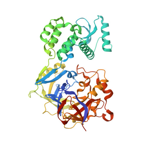
Crystal structure of human mARC1 reveals its exceptional position among eukaryotic molybdenum enzymes.

Kubitza, C.Bittner, F.Ginsel, C.Havemeyer, A.Clement, B.Scheidig, A.J.

(2018) Proc Natl Acad Sci U S A 115: 11958-11963

  • DOI: https://doi.org/10.1073/pnas.1808576115
  • Primary Citation of Related Structures:  
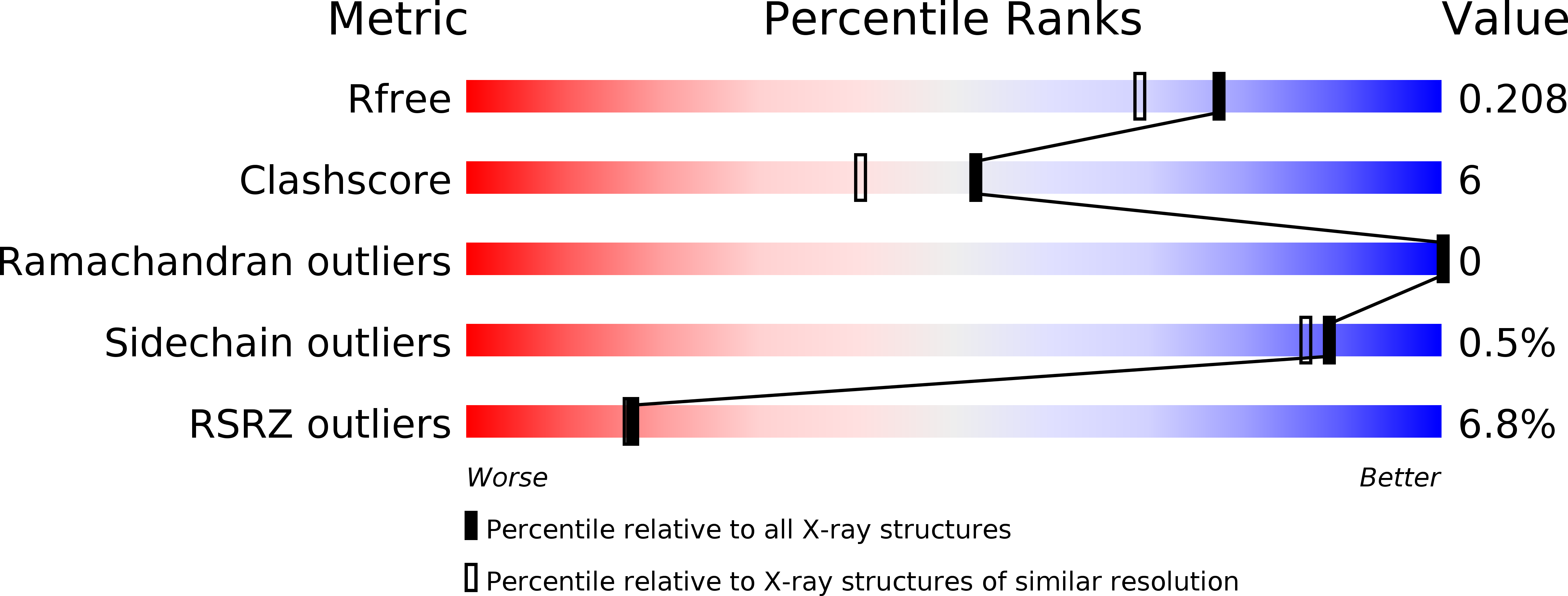
    6FW2

  • PubMed Abstract: 

    Biotransformation enzymes ensure a viable homeostasis by regulating reversible cycles of oxidative and reductive reactions. The metabolism of nitrogen-containing compounds is of high pharmaceutical and toxicological relevance because N-oxygenated metabolites derived from reactions mediated by cytochrome P450 enzymes or flavin-dependent monooxygenases are in some cases highly toxic or mutagenic. The molybdenum-dependent mitochondrial amidoxime-reducing component (mARC) was found to be an extremely efficient counterpart, which is able to reduce the full range of N-oxygenated compounds and thereby mediates detoxification reactions. However, the 3D structure of this enzyme was unknown. Here we present the high-resolution crystal structure of human mARC. We give detailed insight into the coordination of its molybdenum cofactor (Moco), the catalytic mechanism, and its ability to reduce a wide range of N-oxygenated compounds. The identification of two key residues will allow future discrimination between mARC paralogs and ensure correct annotation. Since our structural findings contradict in silico predictions that are currently made by online databases, we propose domain definitions for members of the superfamily of Moco sulfurase C-terminal (MOSC) domain-containing proteins. Furthermore, we present evidence for an evolutionary role of mARC for the emergence of the xanthine oxidase protein superfamily. We anticipate the hereby presented crystal structure to be a starting point for future descriptions of MOSC proteins, which are currently poorly structurally characterized.


  • Organizational Affiliation

    Structural Biology, Zoological Institute, Kiel University, 24118 Kiel, Germany.


Macromolecules
Find similar proteins by:  (by identity cutoff)  |  3D Structure
Entity ID: 1
MoleculeChains Sequence LengthOrganismDetailsImage
Mitochondrial amidoxime-reducing component 1,Endolysin,Mitochondrial amidoxime-reducing component 1459Homo sapiensTequatrovirus T4Mutation(s): 1 
Gene Names: MARC1MOSC1eT4Tp126
EC: 1 (PDB Primary Data), 3.2.1.17 (PDB Primary Data)
UniProt & NIH Common Fund Data Resources
Find proteins for Q5VT66 (Homo sapiens)
Explore Q5VT66 
Go to UniProtKB:  Q5VT66
PHAROS:  Q5VT66
GTEx:  ENSG00000186205 
Find proteins for P00720 (Enterobacteria phage T4)
Explore P00720 
Go to UniProtKB:  P00720
Entity Groups  
Sequence Clusters30% Identity50% Identity70% Identity90% Identity95% Identity100% Identity
UniProt GroupsP00720Q5VT66
Sequence Annotations
Expand
  • Reference Sequence
Small Molecules
Ligands 5 Unique
IDChains Name / Formula / InChI Key2D Diagram3D Interactions
MTE
Query on MTE

Download Ideal Coordinates CCD File 
B [auth A]PHOSPHONIC ACIDMONO-(2-AMINO-5,6-DIMERCAPTO-4-OXO-3,7,8A,9,10,10A-HEXAHYDRO-4H-8-OXA-1,3,9,10-TETRAAZA-ANTHRACEN-7-YLMETHYL)ESTER
C10 H14 N5 O6 P S2
HPEUEJRPDGMIMY-IFQPEPLCSA-N
B3P
Query on B3P

Download Ideal Coordinates CCD File 
D [auth A]2-[3-(2-HYDROXY-1,1-DIHYDROXYMETHYL-ETHYLAMINO)-PROPYLAMINO]-2-HYDROXYMETHYL-PROPANE-1,3-DIOL
C11 H26 N2 O6
HHKZCCWKTZRCCL-UHFFFAOYSA-N
MOO
Query on MOO

Download Ideal Coordinates CCD File 
E [auth A],
F [auth A],
G [auth A],
H [auth A]
MOLYBDATE ION
Mo O4
MEFBJEMVZONFCJ-UHFFFAOYSA-N
EFK
Query on EFK

Download Ideal Coordinates CCD File 
C [auth A]oxidanyl(oxidanylidene)molybdenum
H Mo O2
VEWPYRVWJVBLDN-UHFFFAOYSA-M
PO4
Query on PO4

Download Ideal Coordinates CCD File 
I [auth A]PHOSPHATE ION
O4 P
NBIIXXVUZAFLBC-UHFFFAOYSA-K
Experimental Data & Validation

Experimental Data

  • Method: X-RAY DIFFRACTION
  • Resolution: 1.78 Å
  • R-Value Free: 0.208 
  • R-Value Work: 0.169 
  • R-Value Observed: 0.171 
  • Space Group: P 21 21 21
Unit Cell:
Length ( Å )Angle ( ˚ )
a = 61.133α = 90
b = 74.818β = 90
c = 110.726γ = 90
Software Package:
Software NamePurpose
REFMACrefinement
XDSdata reduction
Aimlessdata scaling
MOLREPphasing
Cootmodel building

Structure Validation

View Full Validation Report


Ligand Structure Quality Assessment 


Entry History 

Deposition Data

Revision History  (Full details and data files)

  • Version 1.0: 2018-10-31
    Type: Initial release
  • Version 1.1: 2018-11-14
    Changes: Data collection, Database references
  • Version 1.2: 2018-11-28
    Changes: Data collection, Database references