6FF9

Mutant R280K of human P53


Experimental Data Snapshot

  • Method: X-RAY DIFFRACTION
  • Resolution: 2.00 Å
  • R-Value Free: 0.226 
  • R-Value Work: 0.182 
  • R-Value Observed: 0.184 

wwPDB Validation   3D Report Full Report


This is version 1.1 of the entry. See complete history


Literature

The Crystal Structure of the R280K Mutant of Human p53 Explains the Loss of DNA Binding.

Gomes, A.S.Trovao, F.Andrade Pinheiro, B.Freire, F.Gomes, S.Oliveira, C.Domingues, L.Romao, M.J.Saraiva, L.Carvalho, A.L.

(2018) Int J Mol Sci 19

  • DOI: https://doi.org/10.3390/ijms19041184
  • Primary Citation of Related Structures:  
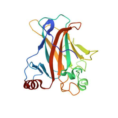
    6FF9

  • PubMed Abstract: 

    The p53 tumor suppressor is widely found to be mutated in human cancer. This protein is regarded as a molecular hub regulating different cell responses, namely cell death. Compelling data have demonstrated that the impairment of p53 activity correlates with tumor development and maintenance. For these reasons, the reactivation of p53 function is regarded as a promising strategy to halt cancer. In the present work, the recombinant mutant p53R280K DNA binding domain (DBD) was produced for the first time, and its crystal structure was determined in the absence of DNA to a resolution of 2.0 Å. The solved structure contains four molecules in the asymmetric unit, four zinc(II) ions, and 336 water molecules. The structure was compared with the wild-type p53 DBD structure, isolated and in complex with DNA. These comparisons contributed to a deeper understanding of the mutant p53R280K structure, as well as the loss of DNA binding related to halted transcriptional activity. The structural information derived may also contribute to the rational design of mutant p53 reactivating molecules with potential application in cancer treatment.


  • Organizational Affiliation

    LAQV-REQUIMTE, Laboratório de Microbiologia, Departamento de Ciências Biológicas, Faculdade de Farmácia, Universidade do Porto, 4050-313 Porto, Portugal. anasarag4@gmail.com.


Macromolecules
Find similar proteins by:  (by identity cutoff)  |  3D Structure
Entity ID: 1
MoleculeChains Sequence LengthOrganismDetailsImage
Cellular tumor antigen p53
A, B, C, D
193Homo sapiensMutation(s): 1 
Gene Names: TP53P53
UniProt & NIH Common Fund Data Resources
Find proteins for P04637 (Homo sapiens)
Explore P04637 
Go to UniProtKB:  P04637
PHAROS:  P04637
GTEx:  ENSG00000141510 
Entity Groups  
Sequence Clusters30% Identity50% Identity70% Identity90% Identity95% Identity100% Identity
UniProt GroupP04637
Sequence Annotations
Expand
  • Reference Sequence
Experimental Data & Validation

Experimental Data

  • Method: X-RAY DIFFRACTION
  • Resolution: 2.00 Å
  • R-Value Free: 0.226 
  • R-Value Work: 0.182 
  • R-Value Observed: 0.184 
  • Space Group: P 1 21 1
Unit Cell:
Length ( Å )Angle ( ˚ )
a = 68.569α = 90
b = 69.445β = 90.04
c = 83.335γ = 90
Software Package:
Software NamePurpose
PHENIXrefinement
XDSdata reduction
Aimlessdata scaling
PHASERphasing
PDB_EXTRACTdata extraction

Structure Validation

View Full Validation Report


Entry History & Funding Information

Deposition Data


Funding OrganizationLocationGrant Number
FCT-MCTESPortugalPTDC/DTP-FTO/1981/2014
FCT-MCTESPortugalPT2020 UID/MULTI/04378/2013
FCT-MCTESPortugalRECI/BBB-BEP/0124/2012

Revision History  (Full details and data files)

  • Version 1.0: 2018-04-25
    Type: Initial release
  • Version 1.1: 2024-01-17
    Changes: Data collection, Database references, Refinement description