6EP9

Crystal structure of BTK kinase domain complexed with N-[2-methyl-3-[4-methyl-6-[4-(4-methylpiperazine-1-carbonyl)anilino]-5-oxo-pyrazin-2-yl]phenyl]-4-(1-piperidyl)benzamide


Experimental Data Snapshot

  • Method: X-RAY DIFFRACTION
  • Resolution: 2.01 Å
  • R-Value Free: 0.239 
  • R-Value Work: 0.199 
  • R-Value Observed: 0.201 

wwPDB Validation   3D Report Full Report


Ligand Structure Quality Assessment 


This is version 1.2 of the entry. See complete history


Literature

Water molecules in protein-ligand interfaces. Evaluation of software tools and SAR comparison.

Nittinger, E.Gibbons, P.Eigenbrot, C.Davies, D.R.Maurer, B.Yu, C.L.Kiefer, J.R.Kuglstatter, A.Murray, J.Ortwine, D.F.Tang, Y.Tsui, V.

(2019) J Comput Aided Mol Des 33: 307-330

  • DOI: https://doi.org/10.1007/s10822-019-00187-y
  • Primary Citation of Related Structures:  
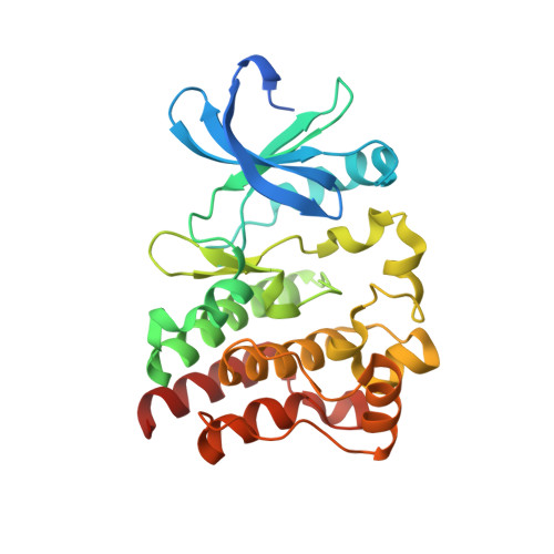
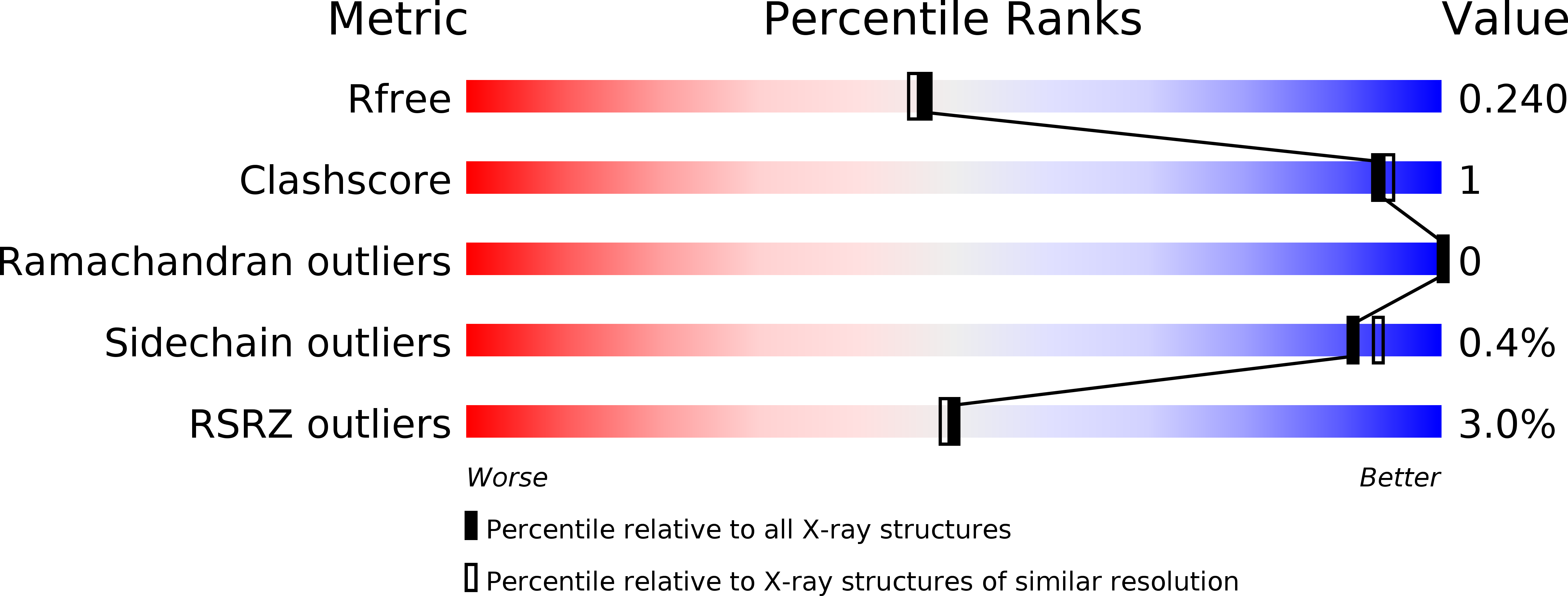
    6BIK, 6BKE, 6BKH, 6BKW, 6BLN, 6BQA, 6BQD, 6EP9

  • PubMed Abstract: 

    Targeting the interaction with or displacement of the 'right' water molecule can significantly increase inhibitor potency in structure-guided drug design. Multiple computational approaches exist to predict which waters should be targeted for displacement to achieve the largest gain in potency. However, the relative success of different methods remains underexplored. Here, we present a comparison of the ability of five water prediction programs (3D-RISM, SZMAP, WaterFLAP, WaterRank, and WaterMap) to predict crystallographic water locations, calculate their binding free energies, and to relate differences in these energies to observed changes in potency. The structural cohort included nine Bruton's Tyrosine Kinase (BTK) structures, and nine bromodomain structures. Each program accurately predicted the locations of most crystallographic water molecules. However, the predicted binding free energies correlated poorly with the observed changes in inhibitor potency when solvent atoms were displaced by chemical changes in closely related compounds.


  • Organizational Affiliation

    Universität Hamburg, ZBH - Center for Bioinformatics, Bundesstraße 43, 20146, Hamburg, Germany. nittinger@zbh.uni-hamburg.de.


Macromolecules
Find similar proteins by:  (by identity cutoff)  |  3D Structure
Entity ID: 1
MoleculeChains Sequence LengthOrganismDetailsImage
Tyrosine-protein kinase BTK283Homo sapiensMutation(s): 4 
Gene Names: BTKAGMX1ATKBPK
EC: 2.7.10.2
UniProt & NIH Common Fund Data Resources
Find proteins for Q06187 (Homo sapiens)
Explore Q06187 
Go to UniProtKB:  Q06187
PHAROS:  Q06187
GTEx:  ENSG00000010671 
Entity Groups  
Sequence Clusters30% Identity50% Identity70% Identity90% Identity95% Identity100% Identity
UniProt GroupQ06187
Sequence Annotations
Expand
  • Reference Sequence
Small Molecules
Ligands 1 Unique
IDChains Name / Formula / InChI Key2D Diagram3D Interactions
BNB (Subject of Investigation/LOI)
Query on BNB

Download Ideal Coordinates CCD File 
B [auth A]N-[2-methyl-3-[4-methyl-6-[4-(4-methylpiperazine-1-carbonyl)anilino]-5-oxo-pyrazin-2-yl]phenyl]-4-(1-piperidyl)benzamide
C36 H41 N7 O3
VDSHGYXLLSEPML-UHFFFAOYSA-N
Experimental Data & Validation

Experimental Data

  • Method: X-RAY DIFFRACTION
  • Resolution: 2.01 Å
  • R-Value Free: 0.239 
  • R-Value Work: 0.199 
  • R-Value Observed: 0.201 
  • Space Group: P 21 21 2
Unit Cell:
Length ( Å )Angle ( ˚ )
a = 72.408α = 90
b = 106.948β = 90
c = 38.126γ = 90
Software Package:
Software NamePurpose
REFMACrefinement
SCALEPACKdata scaling
DENZOdata reduction

Structure Validation

View Full Validation Report


Ligand Structure Quality Assessment 


Entry History 

Deposition Data

Revision History  (Full details and data files)

  • Version 1.0: 2018-08-22
    Type: Initial release
  • Version 1.1: 2019-02-27
    Changes: Data collection, Database references
  • Version 1.2: 2019-03-20
    Changes: Data collection, Database references