6DUK

EGFR with an allosteric inhibitor


Experimental Data Snapshot

  • Method: X-RAY DIFFRACTION
  • Resolution: 2.20 Å
  • R-Value Free: 0.223 
  • R-Value Work: 0.190 
  • R-Value Observed: 0.191 

wwPDB Validation   3D Report Full Report


Ligand Structure Quality Assessment 


This is version 1.3 of the entry. See complete history


Literature

Single and Dual Targeting of Mutant EGFR with an Allosteric Inhibitor.

To, C.Jang, J.Chen, T.Park, E.Mushajiang, M.De Clercq, D.J.H.Xu, M.Wang, S.Cameron, M.D.Heppner, D.E.Shin, B.H.Gero, T.W.Yang, A.Dahlberg, S.E.Wong, K.K.Eck, M.J.Gray, N.S.Janne, P.A.

(2019) Cancer Discov 9: 926-943

  • DOI: https://doi.org/10.1158/2159-8290.CD-18-0903
  • Primary Citation of Related Structures:  
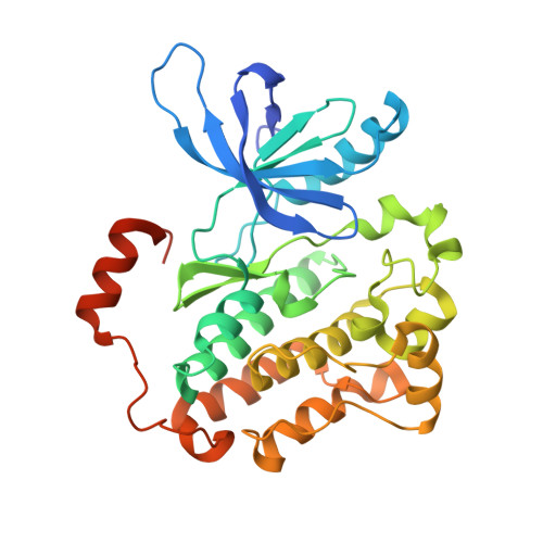
    6DUK

  • PubMed Abstract: 

    Allosteric kinase inhibitors offer a potentially complementary therapeutic strategy to ATP-competitive kinase inhibitors due to their distinct sites of target binding. In this study, we identify and study a mutant-selective EGFR allosteric inhibitor, JBJ-04-125-02, which as a single agent can inhibit cell proliferation and EGFR L858R/T790M/C797S signaling in vitro and in vivo . However, increased EGFR dimer formation limits treatment efficacy and leads to drug resistance. Remarkably, osimertinib, an ATP-competitive covalent EGFR inhibitor, uniquely and significantly enhances the binding of JBJ-04-125-02 for mutant EGFR. The combination of osimertinib and JBJ-04-125-02 results in an increase in apoptosis, a more effective inhibition of cellular growth, and an increased efficacy in vitro and in vivo compared with either single agent alone. Collectively, our findings suggest that the combination of a covalent mutant-selective ATP-competitive inhibitor and an allosteric EGFR inhibitor may be an effective therapeutic approach for patients with EGFR -mutant lung cancer. SIGNIFICANCE: The clinical efficacy of EGFR tyrosine kinase inhibitors (TKI) in EGFR -mutant lung cancer is limited by acquired drug resistance, thus highlighting the need for alternative strategies to inhibit EGFR. Here, we identify a mutant EGFR allosteric inhibitor that is effective as a single agent and in combination with the EGFR TKI osimertinib. This article is highlighted in the In This Issue feature, p. 813 .


  • Organizational Affiliation

    Lowe Center for Thoracic Oncology, Dana-Farber Cancer Institute, Boston, Massachusetts.


Macromolecules
Find similar proteins by:  (by identity cutoff)  |  3D Structure
Entity ID: 1
MoleculeChains Sequence LengthOrganismDetailsImage
Epidermal growth factor receptor
A, B, C, D, E
A, B, C, D, E, F
331Homo sapiensMutation(s): 2 
Gene Names: EGFRERBBERBB1HER1
EC: 2.7.10.1
UniProt & NIH Common Fund Data Resources
Find proteins for P00533 (Homo sapiens)
Explore P00533 
Go to UniProtKB:  P00533
PHAROS:  P00533
GTEx:  ENSG00000146648 
Entity Groups  
Sequence Clusters30% Identity50% Identity70% Identity90% Identity95% Identity100% Identity
UniProt GroupP00533
Sequence Annotations
Expand
  • Reference Sequence
Small Molecules
Ligands 3 Unique
IDChains Name / Formula / InChI Key2D Diagram3D Interactions
JBJ
Query on JBJ

Download Ideal Coordinates CCD File 
I [auth A]
L [auth B]
O [auth C]
R [auth D]
U [auth E]
I [auth A],
L [auth B],
O [auth C],
R [auth D],
U [auth E],
X [auth F]
(2R)-2-(5-fluoro-2-hydroxyphenyl)-2-{1-oxo-6-[4-(piperazin-1-yl)phenyl]-1,3-dihydro-2H-isoindol-2-yl}-N-(1,3-thiazol-2-yl)acetamide
C29 H26 F N5 O3 S
VHQVOTINPRYDAO-AREMUKBSSA-N
ANP
Query on ANP

Download Ideal Coordinates CCD File 
H [auth A]
K [auth B]
N [auth C]
Q [auth D]
T [auth E]
H [auth A],
K [auth B],
N [auth C],
Q [auth D],
T [auth E],
W [auth F]
PHOSPHOAMINOPHOSPHONIC ACID-ADENYLATE ESTER
C10 H17 N6 O12 P3
PVKSNHVPLWYQGJ-KQYNXXCUSA-N
MG
Query on MG

Download Ideal Coordinates CCD File 
G [auth A]
J [auth B]
M [auth C]
P [auth D]
S [auth E]
G [auth A],
J [auth B],
M [auth C],
P [auth D],
S [auth E],
V [auth F]
MAGNESIUM ION
Mg
JLVVSXFLKOJNIY-UHFFFAOYSA-N
Experimental Data & Validation

Experimental Data

  • Method: X-RAY DIFFRACTION
  • Resolution: 2.20 Å
  • R-Value Free: 0.223 
  • R-Value Work: 0.190 
  • R-Value Observed: 0.191 
  • Space Group: P 1
Unit Cell:
Length ( Å )Angle ( ˚ )
a = 57.481α = 70.38
b = 94.711β = 77.46
c = 97.398γ = 79.14
Software Package:
Software NamePurpose
PHENIXrefinement
ABSdata reduction
ABSdata scaling
PHASERphasing

Structure Validation

View Full Validation Report


Ligand Structure Quality Assessment 


Entry History & Funding Information

Deposition Data


Funding OrganizationLocationGrant Number
National Institutes of Health/National Cancer Institute (NIH/NCI)United StatesR01CA201049
National Institutes of Health/National Cancer Institute (NIH/NCI)United StatesP01CA154303
National Institutes of Health/National Cancer Institute (NIH/NCI)United StatesR50CA221830

Revision History  (Full details and data files)

  • Version 1.0: 2019-06-05
    Type: Initial release
  • Version 1.1: 2019-07-17
    Changes: Data collection, Database references
  • Version 1.2: 2019-12-04
    Changes: Author supporting evidence
  • Version 1.3: 2023-10-11
    Changes: Data collection, Database references, Derived calculations, Refinement description